Review
. 2011 May;84(1001):469-77.
doi: 10.1259/bjr/59197814.

Pictorial review of mucopolysaccharidosis with emphasis on MRI features of brain and spine

Affiliations
Review

Pictorial review of mucopolysaccharidosis with emphasis on MRI features of brain and spine

D D Rasalkar et al. Br J Radiol. 2011 May.

Abstract

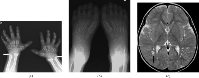
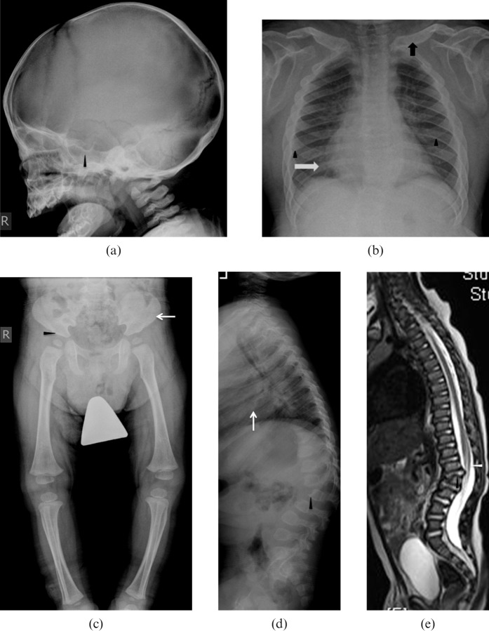
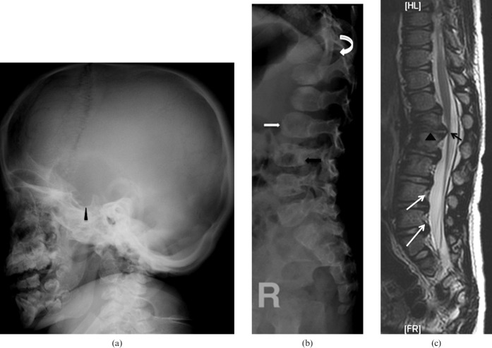
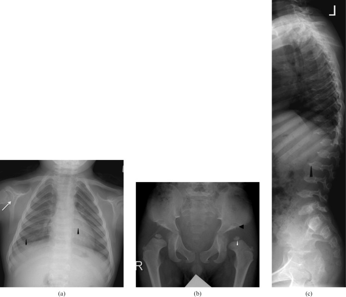
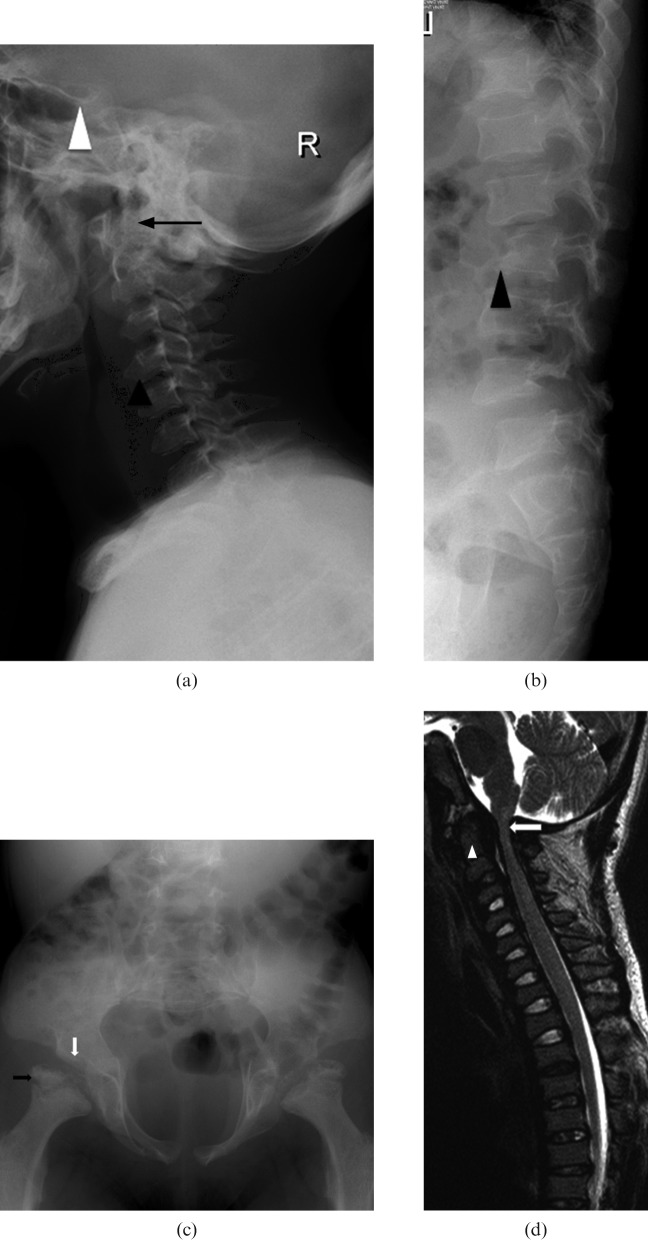
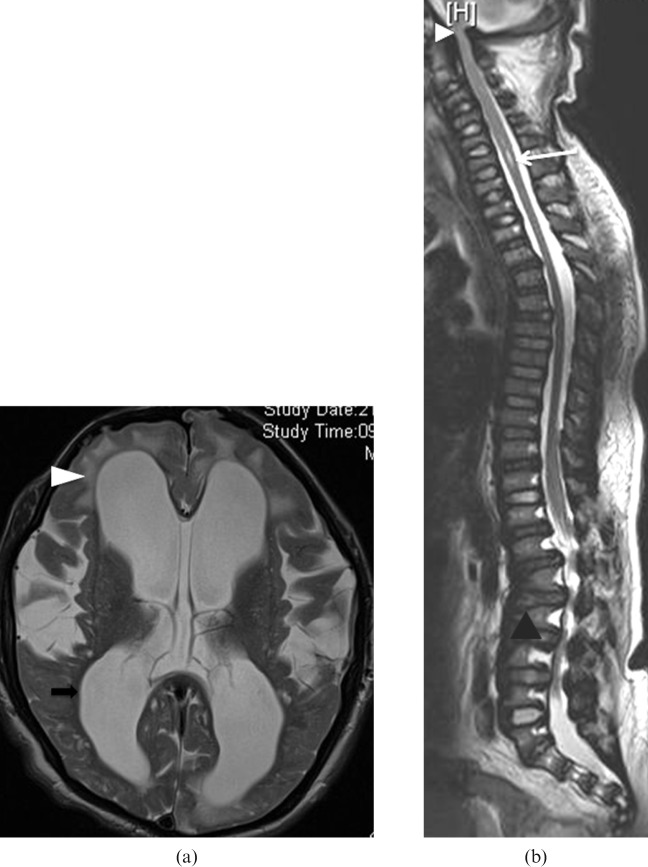
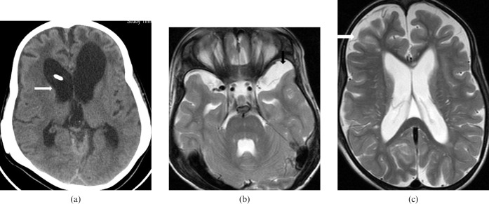
Mucopolysaccharidosis (MPS) is an inherited metabolic disorder of childhood, characterised by progressive multisystem involvement predominantly affecting the skeletal system leading to skeletal dysplasia. Mental retardation, neuropathy and cardiomyopathy may occur in the most severely affected patients, leading to progressive disability and death in their early third to fourth decades. The purpose of this paper is to illustrate the typical imaging features of different types of MPS, in particular the MR features of the brain and spine in MPS, which are expected to be encountered by radiologists more frequently in their clinical practice as a result of prolonged life expectancy for those with MPS with recent advances in therapeutic interventions. The treatment options and outcomes for MPS patients are also briefly discussed.


Figures

Figure 1
Figure 1
Mucopolysaccharidosis (MPS) Type I-S. A 5-year-old female has bilateral calcaneovalgus deformities of the feet, blue sclera and corneal clouding. She is neurologically normal. She has received bone marrow transplantation (BMT) from a human leukocyte antigen (HLA) identical sibling. Her post-BMT course is uneventful with rapid stable engraftment. (a) Anteroposterior (AP) view of wrist and forearm shows classic features of MPS. Note the short and wide metacarpals with proximal pointing (black arrows). Note the Madelung's deformity of the hands with tilting of distal radii and ulna towards each other (white arrows). (b) AP view of feet shows bilateral calcaneovalgus deformities of the feet. (c) MRI brain T2 weighted axial image shows prominent perivascular spaces with cribriform appearance (black arrows). She is neurologically normal with normal intelligence.
Figure 2
Figure 2
Mucopolysaccharide (MPS) Type I. A 2-year-old male has severe lumbar kyphosis but no neurological deficit on examination. He received double unit cord blood transplant. However, post-transplant, the patient developed severe graft vs host disease (GVHD) and died owing to severe immunosuppression and respiratory failure. (a) Lateral view of skull showing classical features of macrocephaly, J-shaped sella turcica (arrowhead) and platyspondyly (white arrows). (b) Frontal radiograph thorax showing cardiomegaly (white arrow), thick ribs (arrow heads) and short thick clavicles (black arrow). (c) Anteroposterior view of pelvis and thighs showing classical features with flattening of acetabuli (arrowhead) with squaring of iliac blades. (d) Lateral view of spine showing wide ribs (white arrow), thoracolumbar kyphosis, anterior beaking in L2 and L3 bodies (arrowhead). (e) Corresponding T2 weighted sagittal image of the spine showing the same acute kyphosis at the L1–2 level, associated with posterior disc herniation (black arrowhead). There is mild narrowing of spinal canal at this level with indentation onto the anterior thecal space but no significant compression of conus medullaris (white arrowhead).
Figure 3
Figure 3
Mucopolysaccharide (MPS) Type II. A 2-year old male presented with bilateral lower limb weakness. He has mild developmental delay and moderate mental retardation. He has received physiotherapy and occupational therapy and is currently in a static course of disease. (a) Lateral radiograph of skull showing scaphocephaly and J-shaped sella turcica (arrowhead). (b) Lateral radiograph of the spine showing central beaking of the vertebrae (white arrow) and posterior vertebral scalloping (black arrow). (c) MRI spine T2 weighted sagittal image showing thoracolumbar kyphosis and gibbus at L1–2 level (arrowheads). Note the mild spinal canal narrowing at L1 and L2 without significant compression of conus medullaris (black arrow). There is also evidence of posterior vertebral scalloping (white arrows).
Figure 4
Figure 4
Mucopolysaccharide (MPS) Type IV. A 6-year-old male has developmental delay and short stature. He has normal intelligence and no neurological deficit. He has received supportive physiotherapy and occupational therapy and is currently in a static course of disease. (a) Frontal chest radiograph shows thick ribs (black arrowhead) and hypoplastic glenoids fossae (white arrow). (b) Anteroposterior radiograph of the pelvis and thighs showing bilateral flared iliac wings, flattened acetabular roofs (black arrowhead) and dysplastic femoral epiphyses (white arrowhead). (c) Lateral radiograph of the spine showing platyspondyly and central beaking of the vertebrae (arrowhead).
Figure 5
Figure 5
Mucopolysaccharide (MPS) Type VI. A 7-year-old boy has pectus excavatum, fixed flexion contractures of knees (with genu varum), elbows, wrists and the interphalangeal joints (with claw hands) and is suffering from progressive bilateral upper and lower limb weakness. He has bilateral corneal cloudiness with impaired vision. He also has recurrent bilateral otitis media with chronic suppurative middle ear effusion and conductive hearing impairment. He has received cord blood transplant (CBT) from a human leukocyte antigen (HLA)-identical younger brother. Subsequent bone marrow examination and DNA study showed satisfactory engraftment. (a) Lateral radiograph of cervical spine showing J-shaped sella (white arrowhead) dysplastic odontoid process (black arrow). Note the anterior beaking of cervical vertebrae (black arrowhead). (b) Lateral radiograph of lumbar spine showing posterior scalloping and hypoplastic L2 vertebral body (arrowhead). Note the Grade I retrolisthesis of L2 over L3. (c) Radiograph of pelvis showing hypoplastic acetabular roofs (white arrow), bilateral femoral subluxations, elongated pelvic inlet with flared iliac wings and fragmented femoral epiphyses (black arrow). (d) MRI spine T2 weighted sagittal image showing dysplastic odontoid process (arrow head), narrowing of the foramina magnum and cervical canal, which is most severe at the craniocervical junctions (white arrow); however, no myelomalacia or syringomyelia is noted.
Figure 6
Figure 6
Mucopolysaccharide (MPS) Type VI. 12-year-old male suffering from repeated convulsions and on tegretol and epilim. Early in his life, the boy was noticed to have a big head, coarse facial features, a flat nasal bridge, protruding eyes, short stature, lumbar lordosis, hearing impairment and progressively increasing bilateral corneal haziness leading to blindness. He also has bilateral inguinal hernia and multiple joint stiffness including wrists, elbows, hips, knees and ankles. He is currently on enzyme replacement therapy. (a) T2 weighted axial image of brain showing moderate hydrocephalus (black arrow), bi-frontal white matter T2 hyperintensities suggestive of bifrontal demyelination (arrowhead). (b) MR spine T2 weighted sagittal image showing flattened vertebral bodies and disc protrusions at multiple levels (black arrowhead). Spinal canal narrowing and cord indentation is noted at the cervicomedullary junction (white arrowhead). Short segment syringomyelia is present at C4–6 levels (white arrow).
Figure 7
Figure 7
Mucopolysaccharide (MPS) Type VI. 7-year-old female, the younger sibling of the case in Figure 6, has short stature, coarse facial features, flat nasal bridge, severe lumbar kyphosis, bilateral corneal haziness, severe joint contractures and bilateral lower limb weakness. She has received peripheral blood stem cell transplant (PBSCT), followed by an uneventful clinical course. (a) CT brain axial image showing dolichocephaly and moderate hydrocephalus (white arrow). (b, c) MRI brain T2 weighted axial images showing left temporal arachnoid cyst (black arrow) and prominent cerebral sulci (white arrow) suggestive of brain atrophy.

References

    1. Morquio L. Sur une forme de dystrophie osseuse familiale. Archives de médicine des infants 1929;32:129–35
    1. Goidanich IF, Lenzi L. Morquio- Ullrich Disease. A new mucopolysaccharidosis. J Bone Joint Surg Am 1964;46:734–46 - PubMed
    1. Langer LO., Jr Spondyloepiphysial dysplasia tarda. Hereditary chondrodysplasia with characteristic vertebral configuration in the adult. Radiology 1964;82:833–9 - PubMed
    1. Langer LO, Jr, Carey LS. The roentgenographic features of the KS mucopolysaccharidosis of Morquio (Morquio-Brailsford's disease). Am J Roentgenol Radium Ther Nucl Med 1966;97:1–20 - PubMed
    1. Kaendler S, Bockenheimer S, Grafin Vitzthum H, Galow W. Cervical myelopathy in mucopolysaccharidosis type II (Hunter's syndrome). Neuroradiologic, clinical and histopathologic findings. Dtsch Med Wochenschr 1990;115:1348–52 - PubMed