. 2011 Jun-Jul;32(6):1021-9.
doi: 10.3174/ajnr.A2463. Epub 2011 Apr 21.

MR imaging findings in patients with secondary intracranial hypertension

Affiliations

MR imaging findings in patients with secondary intracranial hypertension

A C Rohr et al. AJNR Am J Neuroradiol. 2011 Jun-Jul.

Abstract

Background and purpose: IH can alter the configuration of anatomic structures of the central nervous system. We determined the sensitivity and specificity of MR imaging to detect these changes in patients with secondary IH.

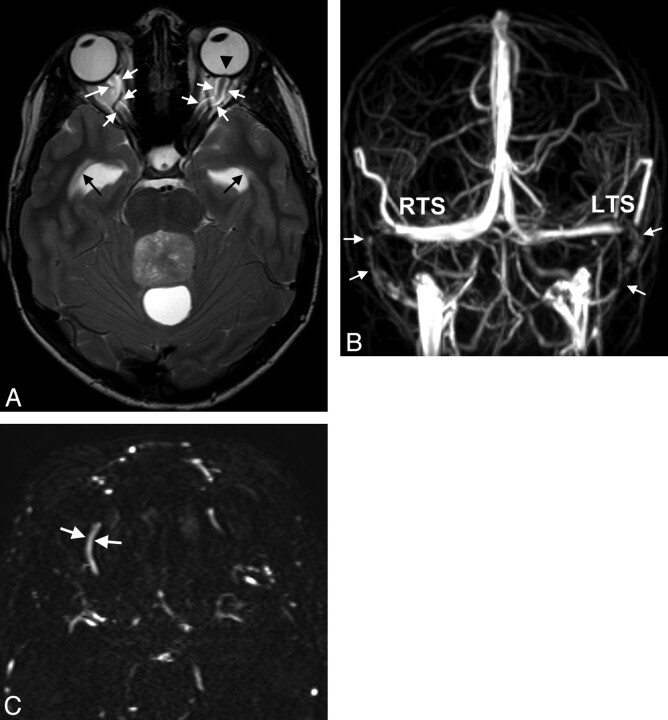
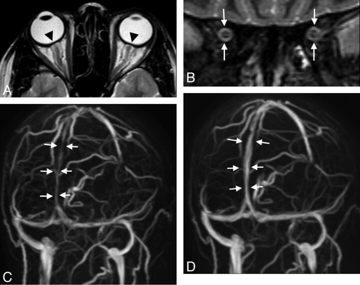
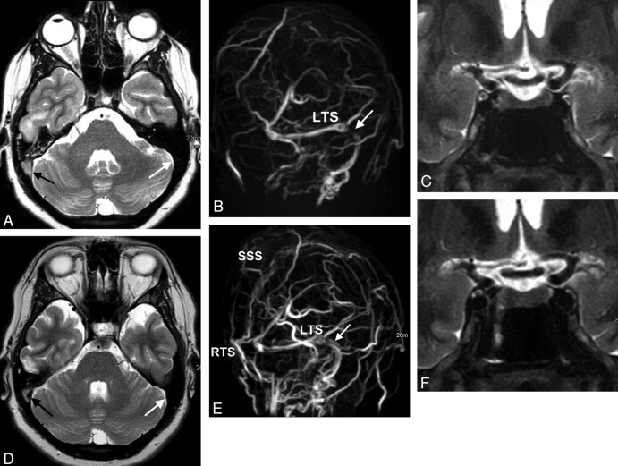
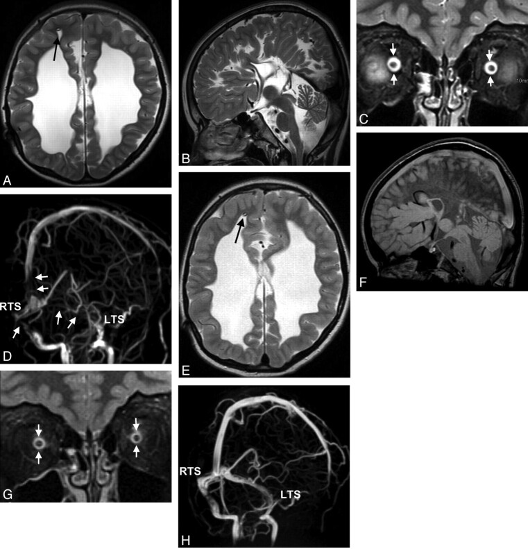
Materials and methods: Patients (n = 36) with IH were prospectively investigated with MR imaging and were matched to 36 controls. MR images were evaluated for elongation and edema of the optic nerves, protrusion of the optic disc, flattening of the posterior sclera, height of the pituitary gland, and width of the optic nerve sheath. On MRV, we recorded venous sinus abnormalities and measured the luminal width of the superior ophthalmic veins. A grading score was introduced to define cranial venous outflow obstruction.

Results: Cranial venous outflow obstruction and ONS hydrops were the most valid signs indicating IH with a sensitivity of 94% and 92% and a specificity of 100% and 89%, respectively. Sensitivities and specificities were 56% and 97% for reduced pituitary height, 64% and 78% for flattening of the posterior sclera, 31% and 97% for widening of the superior ophthalmic veins, 33% and 100% for optic disc protrusion, 14% and 100% for optic nerve edema, and 6% and 100% for elongation of the optic nerve. At least 2 MR imaging findings could be demonstrated in each patient but in none of the controls. The number of positive MR imaging findings correlated with CSF pressure (r = 0.62, P = .01).

Conclusions: The combination of cranial and orbital MR imaging and MRV can be highly sensitive and specific in the diagnosis of patients with IH.


Figures

Fig 1.
Fig 1.
A 15-year-old boy with double vision and reduced acuity has bilateral papilledema. A, On axial T2WI, ventriculomegaly with periventricular CSF flow (black arrows) due to an obstructing tumor within the fourth ventricle is found. Optic nerve sheath hydrops, elongation of the optic nerve (white arrows in A), and optic papilla protrusion are seen (black arrowhead). B, Anteroposterior view of MIP of MRV depicts bilateral narrowings of the lateral RTS and LTS and the sigmoid sinus (arrows). C, On primary axial sections of MRV, the SOVs are enlarged (arrows, right SOV, 2.8-mm width). Medulloblastoma was found at surgery.
Fig 2.
Fig 2.
A 30-year-old woman with arterial hypertension and visual disturbances has bilateral papilledema. A, MR imaging of the orbit displays a flattened posterior sclera (arrowheads). B, ONS hydrops is present, and there is edema of the optic nerve as seen on the coronal STIR sequence measured 20 mm behind the globe (white arrows, ONS width of 5.4 mm). C, Slightly oblique MIP of MRV shows lengthy narrowings of the intracranial venous sinuses, especially of the superior sagittal sinus (arrows). Vision returns to normal with successful treatment of hypertension. On follow-up MR imaging 3 months later, ONS width is reduced to 4.8 mm, and the posterior sclera appears normal (not shown). D, The intracranial venous sinuses regain normal caliber, as shown by MRV.
Fig 3.
Fig 3.
A 28-year-old woman with venous sinus thrombosis secondary to in vitro fertilization. A, Signal intensity typical for thrombosed blood is seen in the SSS and the RTS on axial T2w (black arrow) but not in the LTS (white arrow). B, There is corresponding signal-intensity loss on MIP of MRV in the RTS and SSS (left oblique view). There is a stenosis in the LTS (white arrow) without evidence of a thrombus. C, ONS hydrops is present (not shown), and the height of the pituitary gland is reduced to 2.5 mm (coronal STIR). D and E, CSF pressure is 40 cm H2O. Six months later, partial recanalization of the RTS and SSS occurs following therapy with low-molecular heparin seen on T2WI (black arrow in D) and on MIPs of MRV (E). The stenosis in the LTS is believed to be the result of IH vanishing (white arrow in E). F, The pituitary height-weight returns to normal (4.5 mm).
Fig 4.
Fig 4.
Cranial MR imaging of a 7-year-old boy with congenital posthemorrhagic hydrocephalus before (A–D) and 7 days later after (E–H) correction of the distal part of an insufficient ventriculoperitoneal shunt (arrows in A and E). Standard imaging (axial T2WI in A and E; sagittal T2WI and FLAIR in B and F) does not reflect IH, and does not change after therapy. ONS hydrops (arrows in C, width of 6.8 mm measured in a plane 3 mm dorsal to the optic globe; upper normal limit is 6.3 mm) normalizes after therapy (arrows in G, width of 5.9 mm). Oblique views of MIP of MRV (D and H) display signal-intensity losses in the LTS and RTS and the SSS (arrows in D), also normalizing on follow-up (H). Bilateral papilledema resolves.

References

    1. Ball AK, Clarke CE. Idiopathic intracranial hypertension. Lancet Neurol 2006; 5: 433– 42 - PubMed
    1. Binder DK, Horton JC, Lawton MT, et al. . Idiopathic intracranial hypertension. Neurosurgery 2004; 54: 538– 51, discussion 551–32 - PubMed
    1. Skau M, Brennum J, Gjerris F, et al. . What is new about idiopathic intracranial hypertension? An updated review of mechanism and treatment. Cephalalgia 2006; 26: 384– 99 - PubMed
    1. Bershad EM, Humphreis WE, Suarez JI. Intracranial hypertension. Semin Neurol 2008; 28: 690– 702 - PubMed
    1. Algahtani HA, Baeesa SS, Obeid TH, et al. . Idiopathic intracranial hypertension: atypical presentation. Saudi Med J 2007; 28: 762– 65 - PubMed

Publication types