. 2011 Jun 17;286(24):21384-92.
doi: 10.1074/jbc.M111.241281. Epub 2011 Apr 21.

The G82S polymorphism promotes glycosylation of the receptor for advanced glycation end products (RAGE) at asparagine 81: comparison of wild-type rage with the G82S polymorphic variant

Affiliations

The G82S polymorphism promotes glycosylation of the receptor for advanced glycation end products (RAGE) at asparagine 81: comparison of wild-type rage with the G82S polymorphic variant

Sun Jin Park et al. J Biol Chem. .

Abstract

Interaction between the receptor for advanced glycation end products (RAGE) and its ligands amplifies the proinflammatory response. N-Linked glycosylation of RAGE plays an important role in the regulation of ligand binding. Two potential sites for N-linked glycosylation, at Asn(25) and Asn(81), are implicated, one of which is potentially influenced by a naturally occurring polymorphism that substitutes Gly(82) with Ser. This G82S polymorphic RAGE variant displays increased ligand binding and downstream signaling. We hypothesized that the G82S polymorphism affects RAGE glycosylation and thereby affects ligand binding. WT or various mutant forms of RAGE protein, including N25Q, N81Q, N25Q/G82S, and N25Q/N81Q, were produced by transfecting HEK293 cells. The glycosylation patterns of expressed proteins were compared. Enzymatic deglycosylation showed that WT RAGE and the G82S polymorphic variant are glycosylated to the same extent. Our data also revealed N-linked glycosylation of N25Q and N81Q mutants, suggesting that both Asn(25) and Asn(81) can be utilized for N-linked glycosylation. Using mass spectrometry analysis, we found that Asn(81) may or may not be glycosylated in WT RAGE, whereas in G82S RAGE, Asn(81) is always glycosylated. Furthermore, RAGE binding to S100B ligand is affected by Asn(81) glycosylation, with consequences for NF-κB activation. Therefore, the G82S polymorphism promotes N-linked glycosylation of Asn(81), which has implications for the structure of the ligand binding region of RAGE and might explain the enhanced function associated with the G82S polymorphic RAGE variant.


Figures
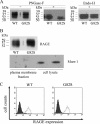
FIGURE 1.
FIGURE 1.
Expression of WT and G82S RAGE on the surface of transiently transfected HEK293 cells. WT and G82S full-length RAGE proteins were expressed by transient transfected HEK293 cells. A, cell lysates containing WT or G82S RAGE without treatment (−) or after PNGase-F or endo-H treatment (+) were subjected to Western blotting, then detected with RAGE-specific antibody. Untreated WT and G82S RAGE preparations comprise three protein species with molecular masses of 59, 57, and 52 kDa. Following PNGase-F treatment, 52-kDa and 50-kDa protein species were detected in WT and G82S RAGE preparations. A PNGase-F-resistant protein band with a molecular mass of 57 kDa was observed only in WT RAGE preparation. Endo-H treatment revealed 57-, 52-, and 50-kDa protein species in both WT and G82S RAGE preparations. B, cell surface biotinylation of RAGE-expressing HEK293 cells revealed that all of the WT and G82S RAGE protein species were expressed on the cell surface. Cell surface proteins were biotinylated and membrane fractions precipitated with NeutrAvidin beads before detection by Western blotting using anti-human RAGE antibody. Endogenous, intracellular protein Murr-1 was detected only in cell lysate preparations. C, cell surface expression of WT and G82S RAGE on HEK293 cells was confirmed by flow cytometry analysis using RAGE-specific antibody and Alexa Fluor 488 rabbit anti-goat antibody.
FIGURE 2.
FIGURE 2.
Comparison of molecular masses of WT and mutant forms (G82S, N25Q, N81Q, N25Q/G82S, N25Q/N81Q) of RAGE. WT and mutant forms of RAGE were expressed in transient transfected HEK293 cells. Cell surface proteins were biotinylated and isolated with NeutrAvidin beads. RAGE proteins in cell lysates and biotinylated RAGE in membrane fraction preparations were detected by Western blotting using RAGE-specific antibody. Endogenous intracellular protein, Murr-1, was detected only in cell lysate preparations. Molecular masses of WT and mutant forms of RAGE were established and are summarized in Table 1.
FIGURE 3.
FIGURE 3.
Extracted ion chromatograms of tryptic peptides containing Asn81 of WT and G82S RAGE. WT and G82S RAGE without treatment or after PNGase-F treatment were digested with trypsin and analyzed by nanoflow HPLC-coupled LTQ Orbitrap MS/MS. Peak intensities of the tryptic peptides from WT (VLPN81GSLFLPAVGIQDEGIFR) and G82S (VLPN81SSLFLPAVGIQDEGIFR) RAGE were plotted as extracted ion chromatograms covering the whole isotope envelopes of both the unmodified (Asn81) and deamidated (Asp81) peptide species. The unmodified peptide is chromatographically separated from the deamidated species with the latter eluting approximately 15 s later. Deglycosylation by PNGase-F increases peak intensities of the deamidated versus unmodified species of the peptides. In G82S only the deamidated form could be detected and only after PNGase-F treatment, indicating that the polymorphic variant is fully glycosylated at Asn81.
FIGURE 4.
FIGURE 4.
S100B-induced, NF-κB p65 activation in RAGE-transfected HEK293 cells. Transient transfected HEK293 cells expressing WT or various mutant forms of RAGE were exposed to S100B (S) or PBS (P). Negative control cells were transfected with pcDNA3.1 (mock). A, nuclear extracts from transfected cells were analyzed by Western blotting using antibodies specific for NF-κB p65, actin, or tubulin as indicated. Increased levels of nuclear NF-κB p65 expression following S100B treatment indicate NF-κB activation. For individual forms of RAGE, the levels of NF-κB p65 expression were variably affected by S100B treatment. The presence of actin (42 kDa) but not tubulin (51 kDa) indicates that nuclear samples were not contaminated by cytosolic proteins. Results are representative of three independent experiments. B, quantitative measures of NF-κB activation in RAGE-transfected cells. Values for the nuclear presence of NF-κB p65 in each sample were normalized relative to nuclear actin. The figure shows mean values (n = 3) for the ratio of normalized nuclear NF-κB p65 present in S100B-treated cells to normalized nuclear NF-κB p65 present in PBS control-treated cells. Statistical comparisons with the same ratio in mock-transfected cells are shown (one-way ANOVA; *, p < 0.05). S100B treatment induced significant NF-κB p65 activation in transfected cells expressing WT and G82S RAGE, as well as N81Q or N25Q/G82S mutant forms of RAGE. Error bars, S.E.
FIGURE 5.
FIGURE 5.
Tertiary structure of RAGE. Figure shows surface molecular structure of the ligand binding V-domain and C1-domain unit of RAGE, based on published coordinates (Protein Data Base ID code 3O3U) (42). The RAGE V-domain contains a hydrophobic cavity (orange) bordered by cationic residues (yellow) and a flexible region (green). The N-linked glycosylation sites, at Asn25 and Asn81, are also shown (purple). The Asn81 site is located adjacent to the hydrophobic cavity that mediates S100B binding (42), whereas the Asn25 site is located on the opposite side of the RAGE molecule.

References

    1. Neeper M., Schmidt A. M., Brett J., Yan S. D., Wang F., Pan Y. C., Elliston K., Stern D., Shaw A. (1992) J. Biol. Chem. 267, 14998–15004 - PubMed
    1. Schmidt A. M., Hori O., Cao R., Yan S. D., Brett J., Wautier J. L., Ogawa S., Kuwabara K., Matsumoto M., Stern D. (1996) Diabetes 45, S77–80 - PubMed
    1. Hofmann M. A., Drury S., Fu C., Qu W., Taguchi A., Lu Y., Avila C., Kambham N., Bierhaus A., Nawroth P., Neurath M. F., Slattery T., Beach D., McClary J., Nagashima M., Morser J., Stern D., Schmidt A. M. (1999) Cell 97, 889–901 - PubMed
    1. Yan S. D., Chen X., Fu J., Chen M., Zhu H., Roher A., Slattery T., Zhao L., Nagashima M., Morser J., Migheli A., Nawroth P., Stern D., Schmidt A. M. (1996) Nature 382, 685–691 - PubMed
    1. Yan S. D., Fu J., Soto C., Chen X., Zhu H., Al-Mohanna F., Collison K., Zhu A., Stern E., Saido T., Tohyama M., Ogawa S., Roher A., Stern D. (1997) Nature 389, 689–695 - PubMed

Publication types

LinkOut - more resources