. 2011 Jun 1;71(11):3872-80.
doi: 10.1158/0008-5472.CAN-10-4482. Epub 2011 Apr 21.

Mild elevation of body temperature reduces tumor interstitial fluid pressure and hypoxia and enhances efficacy of radiotherapy in murine tumor models

Affiliations

Mild elevation of body temperature reduces tumor interstitial fluid pressure and hypoxia and enhances efficacy of radiotherapy in murine tumor models

Arindam Sen et al. Cancer Res. .

Abstract

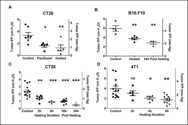
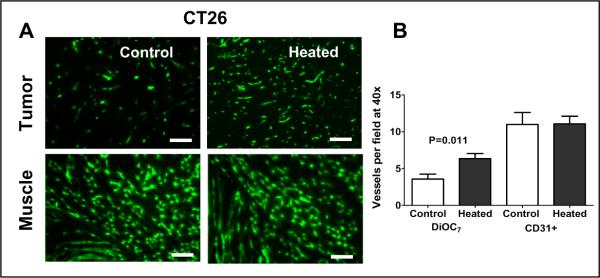
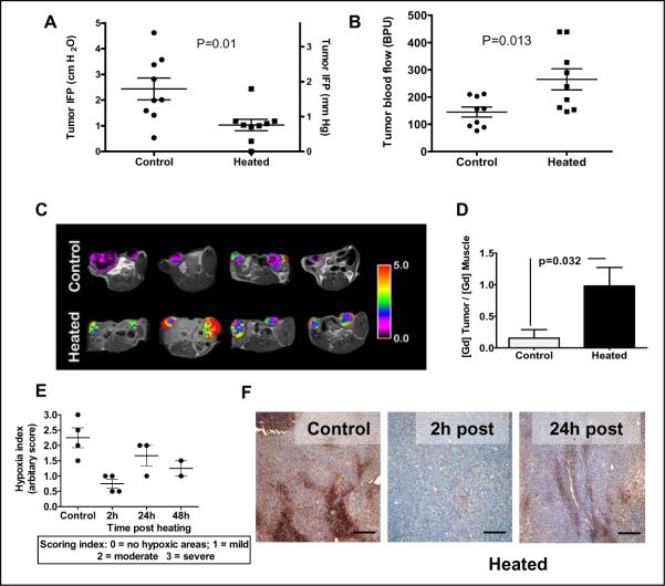
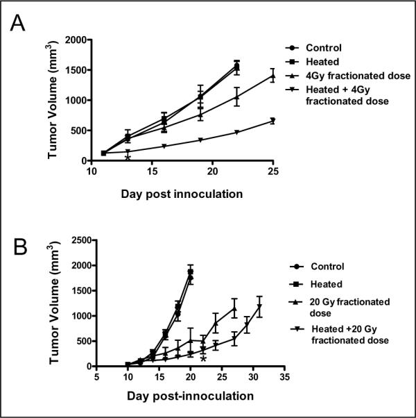
Human and rodent solid tumors often exhibit elevated interstitial fluid pressure (IFP). This condition is recognized as a prognostic indicator for reduced responses to therapy and decreased disease-free survival rate. In the present study, we tested whether induction of a thermoregulatory-mediated increase in tissue blood flow, induced by exposure of mice to mild environmental heat stress, could influence IFP and other vascular parameters within tumors. Using several murine tumor models, we found that heating results in a sustained reduction in tumor IFP correlating with increased tumor vascular perfusion (measured by fluorescent imaging of perfused vessels, laser Doppler flowmetry, and MRI) as well as a sustained reduction in tumor hypoxia. Furthermore, when radiation therapy was administered 24 hours postheating, we observed a significant improvement in efficacy that may be a result of the sustained reduction in tumor hypoxia. These data suggest, for the first time, that environmental manipulation of normal vasomotor function is capable of achieving therapeutically beneficial changes in IFP and microvascular function in the tumor microenvironment.


Figures

Figure 1
Figure 1. An increase in core body temperature decreases tumor interstitial fluid pressures in syngeneic tumors in BALB/c (CT26 and 4T1) and in C57BL/6 (B16.F10) mice
Heated mice bearing tumors (300–600mm3) were maintained at 39.5°C for indicated time, returned to room temperature for 2 hours and then IFP measurements were performed. Each data point represents the average of multiple measurements in a single tumor. (A) CT26 tumors implanted subcutaneously on the hind leg: IFP of tumors in control untreated mice compared to IFP of tumors in mice treated with either 2 doses of paclitaxel (30mg/kg) or heat treatment. (B) B16.F10 tumors implanted subcutaneously on the flank: IFP of tumors in control untreated mice compared to IFP of tumors in mice heated to 39.5°C for 6h. (C) CT26 tumors implanted subcutaneously on the hind leg: IFP of tumors in control mice compared to IFP of tumors in mice after 2h or 4 h heating and 18h and 24h after a 6h heating. (D) 4T1 tumors implanted orthotopically in the 4th mammary fat pad. (300–600mm3). IFP of tumors in control mice and of tumors in mice following 2h, 4h or 6h heating. IFP measurements are given in both cm H2O and mm Hg. ANOVA with Dunnett's multiple comparison was used to test for statistical difference between the mean of the control group and the means of the treated groups (* p,0.05; ** p<0.01; *** p<0.001)
Figure 2
Figure 2. Elevated body temperature increases tumor blood vessel perfusion in BALB/c mice bearing CT26 tumors
(A) Representative fluorescence micrographs of CT26 tumor implanted subcutaneously on the hind leg (300–600mm3 in volume, upper pair) and muscle (lower pair) showing perfused blood vessels following tail vein injection of the cyanine dye DiOC7(3). The micrographs on the left are those obtained from tissue excised from unheated control mice and the two on the right are from mice after 6h heating. The increased number of perfused vessels in a tumor from a heated mouse (160 vessels/this field) is apparent in comparison to that of an untreated mouse (82 vessels/this field). There is no visible difference in the number of perfused vessels in muscles from unheated control and that from heated animals. (B) The average total number of perfused, DiOC7(3) labeled blood vessels in tumors from untheated control mice and those from heated mice showing a doubling in the number of perfused vessels. The average numbers of anatomical CD31+ vessels are not altered by heating. Scale bars represent 500 μm.
Figure 3
Figure 3. Thermally-induced increases in tumor blood flow correlate with decreases in tumor IFP and tissue hypoxia in CT26 tumors
(A) IFP and (B) blood flow changes (as measured by laser Doppler) following heating (n=9/group) of BALB/c mice bearing CT26 tumors implanted subcutaneously on the flanks (300–600mm3 in volume). In these mice, blood flow was measured first and then IFP measurements were taken. (C) Changes in blood flow in CT26 tumors implanted subcutaneously on the hind legs (200–400mm3 in volume) following heating documented by MRI: Representative pseudo-colorized images of contrast agent enhancement in four control animals (top) and another four animals following heating (bottom). Tumors in heated animals consistently showed higher amounts of the contrast agent than tumors in control animals, indicating a higher fractional volume of functioning vasculature as a result of heating. Color scale reflects the ratio of the contrast agent in tumors to normal tissue (back muscle). (D) Plot of the normalized vascular volumes in tumors from unheated control mice and those that heated. Data presented represent the average of each group (control n=8, heated n=10). P-values are Students' T-test results between the control and treated data sets. (D) Plot of the normalized vascular volumes in tumors from unheated control mice and those that heated. Vascular volumes were determined using a macromolecular MR contrast agent HSA-Gd-DTPA. Data presented represent the average of each group (control n=8, heated n=10). P-values are Students' T-test results between the control and the treated data sets. (E) Scatter plot of hypoxia index in tumors (implanted subcutaneously on the flanks, 300–600mm3 in volume) from untreated mice and treated mice at 2, 24 and 48h post heating. (F) Immunohistochemical localization of pimonidazole (Hypoxyprobe-1) adducts in tumors from control mice and from heated mice at 2h and 24h post-heating. A reduction in hypoxic areas in the tumor sections (brown staining) following heating can be seen. Scale bars represent 500 μm.
Figure 4
Figure 4. Heat treatment enhances the efficacy of radiation therapy in two different tumor models
(A) CT26 tumors were implanted subcutaneously on the flank of BALB/c mice. Tumor growth was then monitored in the following groups (n=5 per group): control untreated mice (●), mice that only heated (■), mice that received only radiation (1Gy daily for 4 days, days 12–15; ▲) and mice that heated and followed 24h later by four consecutive radiation treatments (1Gy daily for 4 days, days 12–15; ▼). Tumor growth rates in the unheated control and the heat-only groups were indistinguishable. The slowest growth rate was observed in tumors of mice that received the combination of heating followed by fractionated radiation. Statistical test using oneway ANOVA with Dunnett's multiple comparison tests for the means of the treated groups to the mean of the control group show significant difference only for the group of mice that received combined heating and radiation. (B) B16.F10 tumors were implanted subcutaneously on the flank of C57BL/6 mice. Kinetics of B16.F10 tumor growth in C57BL/6 mice (n=5), control untreated mice (●) and those that were only heated (day 10; ■), only radiation (days 11–15; ▲) and combined radiation and heating (▼). Tumor growth rates in the unheated control and the heating-only groups were comparable. The slowest growth rate was observed in tumors of mice that received fractionated 20Gy radiation (4Gy/day) starting one day after heating. Tumor volumes in the combined heat and radiation treatment group were significantly smaller (P<0.05) as compared to the radiation only group from day 24.

References

    1. Jain RK. Barriers to drug delivery in solid tumors. Sci Am. 1994;271(1):58–65. - PubMed
    1. Dvorak HF. How Tumors Make Bad Blood Vessels and Stroma. Am J Pathol. 2003;162(6):1747–57. - PMC - PubMed
    1. Vaupel P. Tumor microenvironmental physiology and its implications for radiation oncology. Semin Radiat Oncol. 2004;14(3):198–206. - PubMed
    1. Stohrer M, Boucher Y, Stangassinger M, Jain RK. Oncotic pressure in solid tumors is elevated. Cancer Res. 2000;60(15):4251–5. - PubMed
    1. Heldin CH, Rubin K, Pietras K, Ostman A. High interstitial fluid pressure - An obstacle in cancer therapy. Nature Reviews Cancer. 2004;4(10):806–13. - PubMed

Publication types

MeSH terms