. 2011 Aug 1;184(3):317-27.
doi: 10.1164/rccm.201010-1629OC. Epub 2011 Apr 21.

Bronchial secretory immunoglobulin a deficiency correlates with airway inflammation and progression of chronic obstructive pulmonary disease

Affiliations

Bronchial secretory immunoglobulin a deficiency correlates with airway inflammation and progression of chronic obstructive pulmonary disease

Vasiliy V Polosukhin et al. Am J Respir Crit Care Med. .

Abstract

Rationale: Although airway inflammation can persist for years after smoking cessation in patients with chronic obstructive pulmonary disease (COPD), the mechanisms of persistent inflammation are largely unknown.

Objectives: We investigated relationships between bronchial epithelial remodeling, polymeric immunoglobulin receptor (pIgR) expression, secretory IgA (SIgA), airway inflammation, and mural remodeling in COPD.

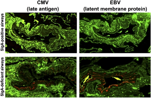
Methods: Lung tissue specimens and bronchoalveolar lavage were obtained from lifetime nonsmokers and former smokers with or without COPD. Epithelial structural changes were quantified by morphometric analysis. Expression of pIgR was determined by immunostaining and real-time polymerase chain reaction. Immunohistochemistry was performed for IgA, CD4 and CD8 lymphocytes, and cytomegalovirus and Epstein-Barr virus antigens. Total IgA and SIgA were measured by ELISA and IgA transcytosis was studied using cultured human bronchial epithelial cells.

Measurements and main results: Areas of bronchial mucosa covered by normal pseudostratified ciliated epithelium were characterized by pIgR expression with SIgA present on the mucosal surface. In contrast, areas of bronchial epithelial remodeling had reduced pIgR expression, localized SIgA deficiency, and increased CD4(+) and CD8(+) lymphocyte infiltration. In small airways (<2 mm), these changes were associated with presence of herpesvirus antigens, airway wall remodeling, and airflow limitation in patients with COPD. Patients with COPD had reduced SIgA in bronchoalveolar lavage. Air-liquid interface epithelial cell cultures revealed that complete epithelial differentiation was required for normal pIgR expression and IgA transcytosis.

Conclusions: Our findings indicate that epithelial structural abnormalities lead to localized SIgA deficiency in COPD airways. Impaired mucosal immunity may contribute to persistent airway inflammation and progressive airway remodeling in COPD.


Figures

Figure 1.
Figure 1.
Structural remodeling of large airway epithelium in chronic obstructive pulmonary disease (COPD) (18). Rows I and II: (A) Pseudostratified ciliated bronchial epithelium with normal ratio of basal, ciliated, and goblet cells. (B) Pseudostratified bronchial epithelium with deviated cell differentiation (goblet cell hyperplasia) showing predomination of goblet cells among scattered ciliated cells. (C) Pseudostratified bronchial epithelium with incomplete cell differentiation; surface cells have columnar shape but do not show morphologic features of ciliated or goblet cells. (D) Stratified bronchial epithelium with incomplete cell differentiation (immature squamous metaplasia); surface cells have cuboid shape and do not contact basement membrane. (E) Bronchial epithelium with altered cell differentiation (complete squamous metaplasia). Row III: Abundant polymeric immunoglobulin receptor (pIgR) (red) is present within normal-appearing pseudostratified epithelium (A), but is reduced in goblet cell hyperplasia (B) and absent in epithelia with incomplete and altered cell differentiation (CE). Reduced pIgR expression in bronchial epithelium correlates with significant reduction in the amount of IgA (green) on the epithelial surface (AE). Row I, hematoxylin and eosin–stained paraffin sections (original magnification ×300); row II, electron micrographs (original magnification ×3,000); row III, double immunofluorescence with anti-pIgR (red) and anti-IgA (green) antibodies, confocal microscope images (original magnification ×400). (F) Distribution of epithelial types shown in A–E lining bronchial mucosal surfaces from lifelong nonsmokers (never smokers), former smokers without COPD, and patients with mild-moderate (Global Initiative for Chronic Obstructive Lung Disease [GOLD] stage I–II) or severe-to-very-severe (GOLD stage III–IV) COPD. Mean ± SEM is indicated for each epithelial subtype. * P < 0.01 compared with never smokers and former smokers without COPD; ** Epithelial disorders not detected in never smokers and former smokers without COPD. Increase in intraepithelial and subepithelial CD8+ lymphocytes (G) or CD4+ lymphocytes (H) with progression of bronchial epithelial structural disorders. Deviated COPD = cell count in areas with deviated epithelial cell differentiation in patients with COPD; Inc-Alt COPD = cell count in areas with incomplete or altered epithelial cell differentiation in patients with COPD; Norm-app COPD = cell count in areas with normal-appearing epithelium in patients with COPD; Norm-app NS = cell count for nonsmokers (both lifelong nonsmokers and former smokers without COPD). Mean ± SEM is indicated for each structural variant. * P < 0.01 compared with nonsmoker group.
Figure 2.
Figure 2.
Small airway mucosal secretory immunoglobulin A (SIgA) deficiency. (A) Airway with normal-appearing respiratory epithelium showing polymeric Ig receptor (pIgR) expression (red) in epithelial cells and surface IgA (green). (B) Focal goblet cell metaplasia showing pIgR expression (red) in epithelial cells and surface SIgA expression (green). (C) Extensive goblet cell metaplasia with profound reduction in pIgR in remaining ciliated cells (red) and absence of surface SIgA (green). (D) Stratified epithelium negative for pIgR and surface SIgA. Row I, hematoxylin and eosin (HE)–stained sections (original magnification ×100); row II, periodic acid–Schiff (PAS)–stained tissue sections (original magnification ×400); row III, immunofluorescence with anti-IgA antibody (green), confocal microscope images (original magnification ×100); row IV, double immunofluorescence with anti-pIgR (red) and anti-IgA (green) antibodies, confocal microscope images (original magnification ×400). (E) Distribution of epithelial types shown in 2A–2D lining small airway mucosa according to clinical status. Mean ± SEM is indicated for each epithelial subtype. * P < 0.01 compared with lifelong non-smokers (never smokers); ** Epithelial disorders not detected in never smokers and former smokers without chronic obstructive pulmonary disease (COPD). (F) Progressive reduction of IgA-specific fluorescent signal on epithelial surfaces of small airways in lifelong non-smokers (never smokers), former smokers without COPD, and patients with COPD. Mean ± SEM is indicated for each clinical group. * P < 0.01 compared with never smokers. (G) Correlation between small airway surface SIgA (estimated by IgA-specific fluorescent signal on epithelial surface) and airflow estimated by FEV1 parameter in former smokers and patients with Grade I–II or Grade III–IV COPD. (H) PIGR mRNA expression in airway epithelial cells. Mean ± SEM is indicated for each clinical group. * P < 0.01 compared with never smokers. Apv = actual pixel value.
Figure 3.
Figure 3.
Association of secretory immunoglobulin A (SIgA) deficiency and herpesvirus infection in small airways. Images of infected and uninfected airways are from the same patient with chronic obstructive pulmonary disease. Double immunofluorescence with primary anti-IgA (green) and anti–Epstein-Barr virus (EBV) or anti-cytomegalovirus (CMV) antibodies (red), confocal microscope images (original magnification ×100).
Figure 4.
Figure 4.
Surface secretory immunoglobulin A (SIgA) deficiency is associated with increased CD8+ T lymphocyte accumulation in small airways. (A) Normal-appearing surface IgA-positive small airway in lifelong nonsmoker with few adjacent CD8+ cells. (B) Normal-appearing surface IgA-positive small airway in a patient with chronic obstructive pulmonary disease (COPD) with slight increase in intraepithelial and submucosal CD8+ cells. (C) Surface IgA-deficient small airway in a patient with COPD with a significant increase in intraepithelial and submucosal CD8+ cells. (AC) Double immunofluorescence with primary anti-IgA (green) and anti-CD8 (red) antibodies, confocal microscope images (original magnification ×100). (D) CD8+ cell accumulation predominates in IgA-deficient airways, leading to increased weighted averages. Mean ± SEM is indicated for IgA-positive and IgA-deficient airways and weighted average in each clinical group. * P < 0.01 compared with lifelong nonsmokers (never smokers). ** P < 0.01 compared with surface IgA-positive airways from the respective clinical group. (E) Inverse correlation between small airway surface IgA (estimated by IgA-specific fluorescent signal on epithelial surface) and airway-associated CD8+ cells. Apv = actual pixel value.
Figure 5.
Figure 5.
Surface secretory immunoglobulin A (SIgA) deficiency is associated with mural remodeling of small airways. (A) Normal-appearing surface SIgA-positive small airway in lifelong nonsmoker showing a thin, nonfibrotic submucosa. (B) Normal-appearing surface SIgA-positive small airway in a patient with chronic obstructive pulmonary disease (COPD) showing moderate submucosal fibrosis, but without significantly increased wall thickness. (C) Surface SIgA-deficient small airway in COPD patient showing marked submucosal fibrosis and significant thickening of the airway wall. Row I, immunofluorescence with anti-IgA antibody (green) (original magnification ×100); red lines delineate airway wall thickness. Row II, trichrome-stained tissue sections (original magnification ×100). (D) Wall thickness (VVsub) predominates in SIgA-deficient airways, leading to increased weighted averages. Mean ± SEM is indicated for SIgA-positive and SIgA-deficient airways and weighted average in each clinical group. * P < 0.01 compared with lifelong nonsmokers (never smokers). ** P < 0.01 compared with surface SIgA-positive airways from the same clinical group. (E) Inverse correlation between small airway surface SIgA (estimated by IgA-specific fluorescent signal on epithelial surface) and airway wall thickness (VVsub). Apv = actual pixel value.
Figure 6.
Figure 6.
Determination of immunoglobulin A (IgA) and secretory IgA (SIgA) in bronchoalveolar lavage (BAL) fluid. (A) Concentration of total IgA and SIgA in BAL fluid from lifelong nonsmokers and former smokers with or without chronic obstructive pulmonary disease. Mean ± SEM is indicated for each clinical group.* P < 0.05 compared with never smokers. (B) Correlation between SIgA concentration in BAL fluid and FEV1. COPD = chronic obstructive pulmonary disease.
Figure 7.
Figure 7.
Polymeric immunoglobulin receptor (pIgR) expression and IgA transcytosis in air-liquid interface (ALI) cultured human bronchial epithelial cells (HBECs) grown with or without retinoic acid (RA). (A) Pseudostratified ciliated structure of bronchial epithelium and marked secretory component (SC)/pIgR expression in culture with RA added; squamous stratified structure of bronchial epithelium and absent SC/pIgR expression in culture grown without RA. Top row: hematoxylin and eosin (HE)–stained paraffin sections (original magnification ×400). Bottom row: immunohistochemistry of paraffin tissue sections with goat polyclonal anti–SC-pIgR antibody (original magnification ×400). (B) Western blot showing expression of SC/pIgR in cell cultures with RA added and no pIgR expression in cells grown without RA. (C) PIGR mRNA expression in RA+ and RA cultures (normalized to HPRT). (D) IgA and secretory IgA (SIgA) concentrations in apical washings of ALI cultured HBECs grown with or without RA. * P < 0.001 (compared with RA+ cultures). Mean ± SEM is indicated for each concentration.

Comment in

References

    1. Rutgers SR, Postma DS, ten Hacken NH, Kauffman HF, Der Mark TW, Koeter GH, Timens W. Ongoing airway inflammation in patients with COPD who do not currently smoke. Thorax 2000;55:12–18 - PubMed
    1. Lapperre TS, Postma DS, Gosman MM, Snoeck-Stroband JB, ten Hacken NH, Hiemstra PS, Timens W, Sterk PJ, Mauad T. Relation between duration of smoking cessation and bronchial inflammation in COPD. Thorax 2006;61:115–121 - PMC - PubMed
    1. Verra F, Escudier E, Lebargy F, Bernaudin JF, De CH, Bignon J. Ciliary abnormalities in bronchial epithelium of smokers, ex-smokers, and nonsmokers. Am J Respir Crit Care Med 1995;151:630–634 - PubMed
    1. Wistuba II, Lam S, Behrens C, Virmani AK, Fong KM, LeRiche J, Samet JM, Srivastava S, Minna JD, Gazdar AF. Molecular damage in the bronchial epithelium of current and former smokers. J Natl Cancer Inst 1997;89:1366–1373 - PMC - PubMed
    1. Lapperre TS, Sont JK, van Schadewijk A, Gosman MM, Postma DS, Bajema IM, Timens W, Mauad T, Hiemstra PS. Smoking cessation and bronchial epithelial remodelling in COPD: a cross-sectional study. Respir Res 2007;8:85. - PMC - PubMed

Publication types

MeSH terms

Substances