Review
. 2011:34:601-28.
doi: 10.1146/annurev-neuro-060909-153200.

General anesthesia and altered states of arousal: a systems neuroscience analysis

Affiliations
Review

General anesthesia and altered states of arousal: a systems neuroscience analysis

Emery N Brown et al. Annu Rev Neurosci. 2011.

Abstract

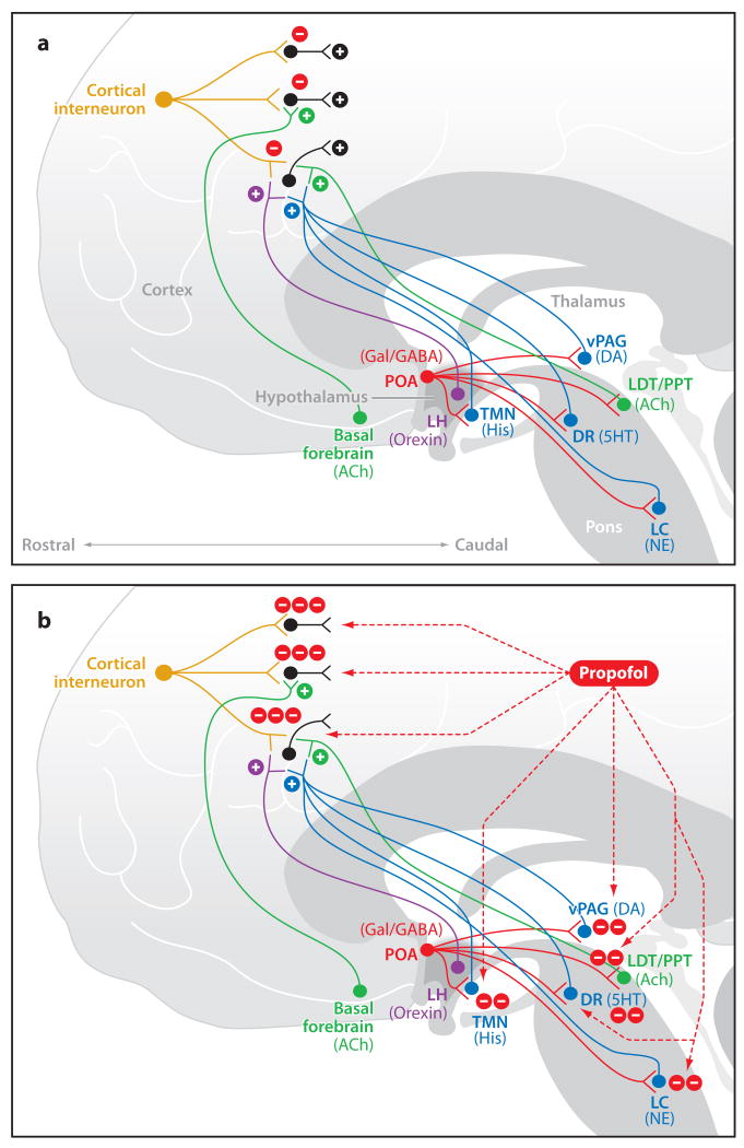
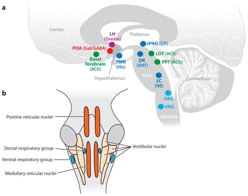
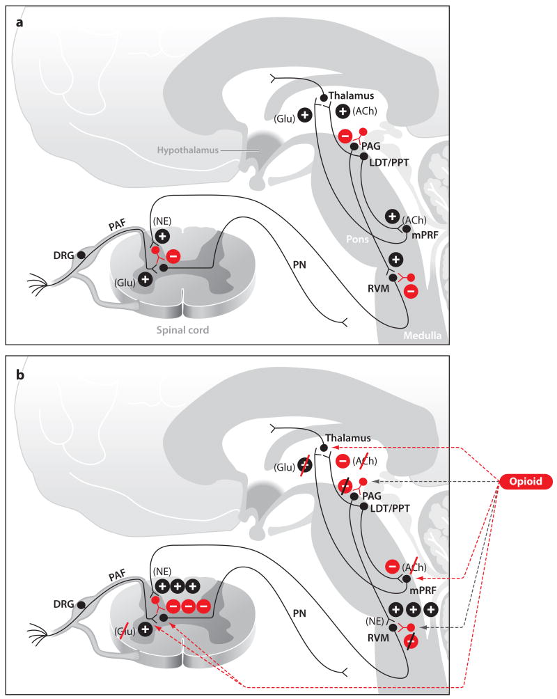
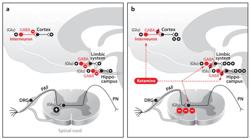
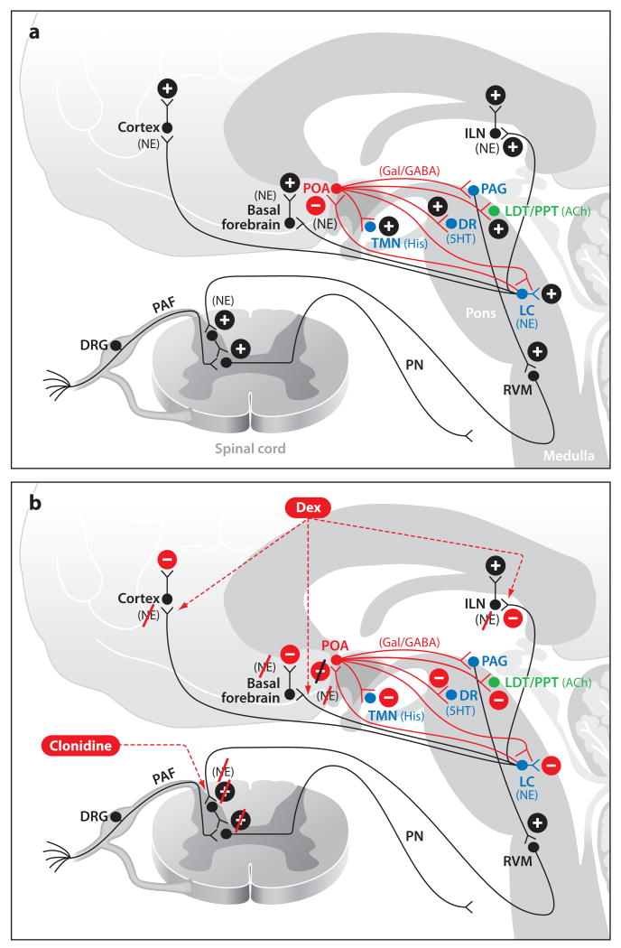
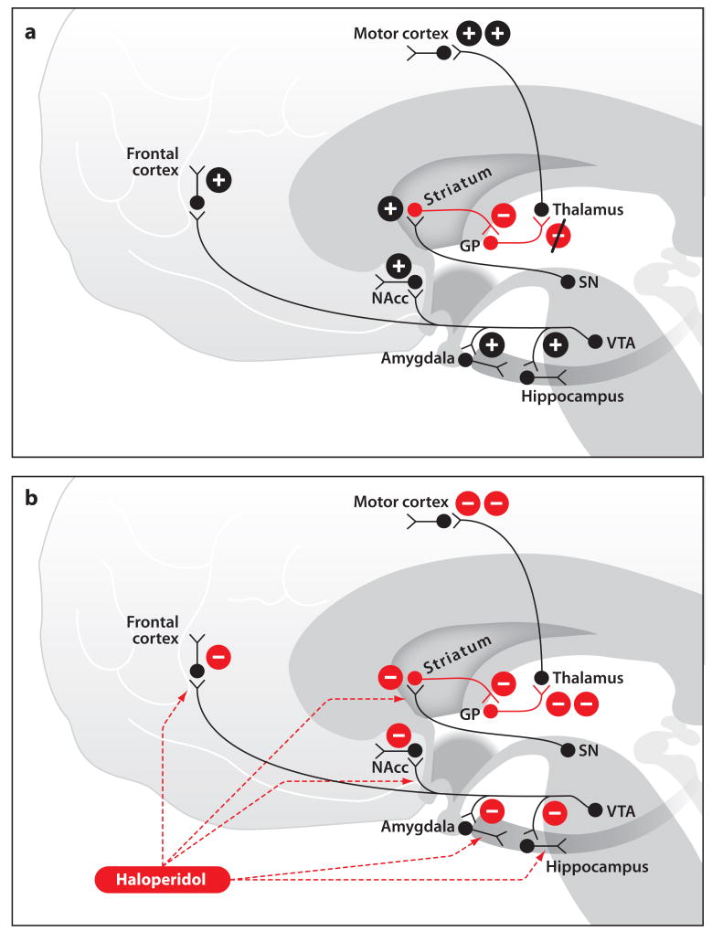
Placing a patient in a state of general anesthesia is crucial for safely and humanely performing most surgical and many nonsurgical procedures. How anesthetic drugs create the state of general anesthesia is considered a major mystery of modern medicine. Unconsciousness, induced by altered arousal and/or cognition, is perhaps the most fascinating behavioral state of general anesthesia. We perform a systems neuroscience analysis of the altered arousal states induced by five classes of intravenous anesthetics by relating their behavioral and physiological features to the molecular targets and neural circuits at which these drugs are purported to act. The altered states of arousal are sedation-unconsciousness, sedation-analgesia, dissociative anesthesia, pharmacologic non-REM sleep, and neuroleptic anesthesia. Each altered arousal state results from the anesthetic drugs acting at multiple targets in the central nervous system. Our analysis shows that general anesthesia is less mysterious than currently believed.


Figures

Figure 1
Figure 1
GABAergic signaling during the awake state (a) and during propofol administration (b). (a) A cortical interneuron during wakefulness mediating control of pyramidal neurons and being modulated by ascending arousal centers. Also shown are inhibitory projections from the POA to the arousal-promoting nuclei. (b) Propofol enhances GABAergic transmission in the cortex and at the inhibitory projections from the POA to the arousal centers. Abbreviations: 5HT, serotonin; ACh, acetylcholine; DA, dopamine; DR, dorsal raphe; GABA, gamma aminobutyric acid; Gal, galanin; His, histamine; LC, locus coeruleus; LDT, laterodorsal tegmental area; LH, lateral hypothalamus; NE, norepinephrine; POA, preoptic area; PPT, pedunculopontine tegmental area; TMN, tuberomamillary nucleus; vPAG, ventral periaquaductal gray.
Figure 2
Figure 2
Sites of major nuclei that regulate arousal and respiration. (a) A sagittal diagram of a human brain with cholinergic nuclei (green), monoaminergic nuclei (dark blue), GABAergic and galanergic nuclei (red), orexin nuclei (pink), and the respiratory nuclei (light blue). (b) Sites of major respiratory and motor relay nuclei in the pons and medulla. Abbreviations: 5HT, serotonin; ACh, acetylcholine; DA, dopamine; DRG, dorsal respiratory group; GABA, gamma aminobutyric acid; Gal, galanin; His, histamine; LC, locus coeruleus; LDT, laterodorsal tegmental area; LH, lateral hypothalamus; NE, norepinephrine; POA, preoptic area; PPT, pedunculopontine tegmental area; TMN, tuberomamillary nucleus; VAG, ventral periaquaductal gray; VRG, ventral respiratory group.
Figure 3
Figure 3
Sites of opioid receptor effects during the awake state (a) and during opioid administration (b). (a) Normal cholinergic signaling during wakefulness and normal nociceptive signaling from the spinal cord to brain stem. (b) Illustrates opioid-induced decrease in arousal through a decrease in cholinergic signaling and the mechanisms of opioid-induced analgesia. Abbreviations: ACh, acetylcholine; DRG, dorsal root ganglia; Glu, glutamate; LDT, laterodorsal tegmental area; mPRF, medial pontine reticular formation; NE, norepinephrine; PAF, peripheral afferent fiber; PAG, periaquaductal gray; PN, projection neuron; PPT, pedunculopontine tegmental area; RVM, rostal ventral medulla.
Figure 4
Figure 4
NMDA receptor signaling during the awake state (a) and during ketamine administration (b). (a) Normal glutamatergic regulation of GABAergic interneuron signaling during wakefulness and normal pain pathway signaling. (b) Ketamine blockage of GABAergic interneuron inhibition and ketamine-induced mechanism of analgesia. Abbreviations: DRG, dorsal root ganglia; GABA, gamma aminobutyric acid; Glu, glutamate; PAF, peripheral afferent fiber; PN, projection neuron.
Figure 5
Figure 5
Alpha adrenergic signaling during the awake state (a) and during dexmedetomidine or clonidine administration (b). (a) Normal adrenergic signaling from the LC during wakefulness and normal pain signaling during wakefulness from the spinal cord to the brain stem. (b) Dexmedetomidine-induced loss of consciousness through NE mediated inhibition of the POA and decreased noradrenergic signaling in the thalamus and cortex. Clonidine-induced analgesia through enhanced inhibitory activity in the descending pain pathway. Abbreviations: 5HT, serotonin; ACh, acetylcholine; DA, dopamine; DRG, dorsal root ganglia; GABAA, gamma aminobutyric acid receptor subtype A; Gal, galanin; His, histamine; ILN, intralaminar nucleus of the thalamus; LC, locus coeruleus; LDT, laterodorsal tegmental area; NE, norepinephrine; PAF, peripheral afferent fiber; PN, projection neuron; POA, preoptic area; PPT, pedunculopontine tegmental area; RVM, rostral ventral medulla; TMN, tuberomamillary nucleus; vPAG, ventral periaquaductal gray.
Figure 6
Figure 6
Dopamine signaling during the awake state (a) and during haloperidol administration (b). (a) Normal dopamine signaling during wakefulness. (b) Haloperidol effects on dopamine signaling. Abbreviations: GP, globus pallidus; NAcc, nucleus accumbens; SN, substantia nigra; VTA, ventral tegmental area.

References

    1. Ali HH, Utting JE, Gray TC. Stimulus frequency in the detection of neuromuscular block in humans. Br J Anaesth. 1970;42(11):967–78. - PubMed
    1. Ali HH, Utting JE, Gray TC. Quantitative assessment of residual antidepolarizing block. II. Br J Anaesth. 1971;43(5):478–85. - PubMed
    1. Alkire MT, Haier RJ, Barker SJ, Shah NK, Wu JC, Kao YJ. Cerebral metabolism during propofol anesthesia in humans studied with positron emission tomography. Anesthesiology. 1995;82(2):393–403. discussion 27A. - PubMed
    1. Alkire MT, Hudetz AG, Tononi G. Consciousness and anesthesia. Science. 2008;322(5903):876–80. - PMC - PubMed
    1. Anand KJ. Anesthetic neurotoxicity in newborns: Should we change clinical practice? Anesthesiology. 2007;107(1):2–4. - PubMed

Publication types