. 2011 Aug;17(15-16):2123-32.
doi: 10.1089/ten.TEA.2010.0637.

Characterization of urine-derived stem cells obtained from upper urinary tract for use in cell-based urological tissue engineering

Affiliations

Characterization of urine-derived stem cells obtained from upper urinary tract for use in cell-based urological tissue engineering

Shantaram Bharadwaj et al. Tissue Eng Part A. 2011 Aug.

Abstract

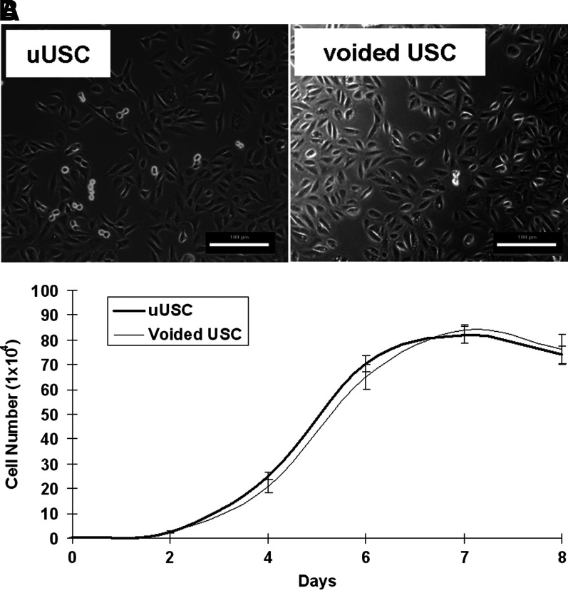
Background: The goals of this study were to characterize urine-derived stem cells obtained from the upper urinary tract (uUSC), induce these cells to differentiate into urothelial and smooth muscle cells, and determine whether they could serve as a potential stem cell source for bladder tissue engineering.

Materials and methods: Urine samples were collected from five patients with normal upper urinary tracts during renal pyeloplasty. Cells were isolated from this urine and extensively expanded in vitro.

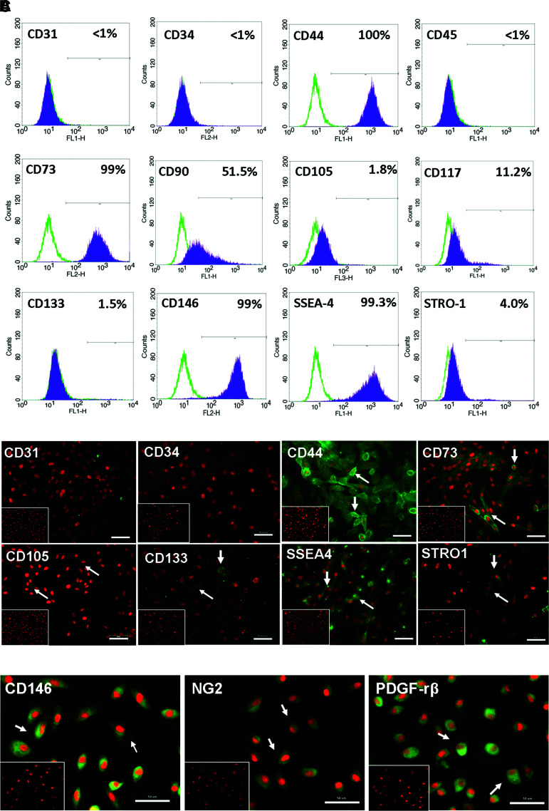
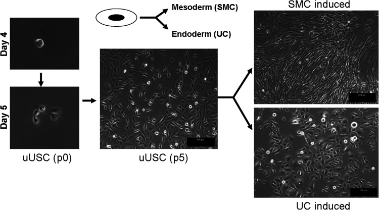
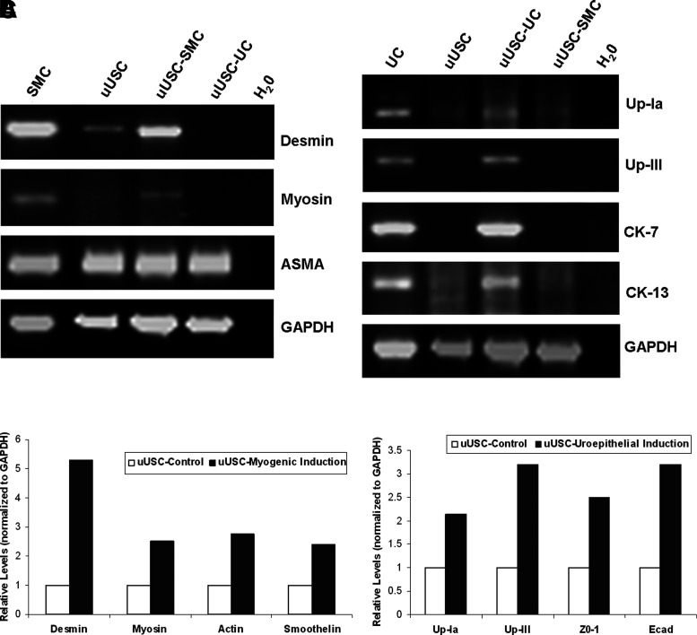
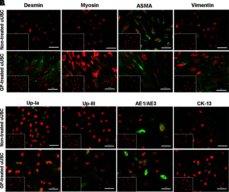
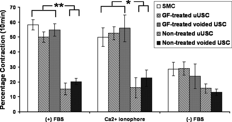
Results: The mean population doubling of uUSC was 46.5±7.7. The uUSC expressed surface markers associated with mesenchymal stem cells and pericytes. These cells could differentiate into smooth muscle-like cells that expressed smooth muscle-specific gene transcripts and proteins, including α-smooth muscle actin, desmin, and myosin, when exposed to TGF-β1 and PDGF-BB. In a collagen lattice assay, these myogenic-differentiated uUSC displayed contractile function that was similar to that seen in native smooth muscle cells. Urothelial-differentiated uUSC expressed urothelial-specific genes and proteins such as uroplakin-Ia and -III, cytokeratin (CK)-7, and CK-13.

Conclusions: uUSC possess expansion and differentiation (urothelial and myogenic) capabilities, and can potentially be used as an alternative cell source in bladder tissue engineering for patients needing cystoplasty.


Conflict of interest statement

No competing financial interests exist.

Figures

FIG. 1.
FIG. 1.
Morphology and growth of uUSC. (A) A single clone of uUSC shown along with voided USC at passage 2 demonstrating the similarity in morphology of the two cell clones. Scale bar=100 μm. (B) The growth pattern of uUSC was similar to voided USC at passage 5. The average doubling time for both the cells was around 20 h. uUSC, urine-derived stem cells obtained from the upper urinary tract.
FIG. 2.
FIG. 2.
(A) Analysis of surface marker expression in uUSC by FACS. A clone of uUSC (p4) was stained with CD markers expressed by mesenchymal as well as hematopeietic stem cells. All MSC markers stained positive, whereas the hematopoitic markers (CD31, 34, and 45) were negative. (B) Immunofluorescence staining for stem cell markers. Immunofluorescent images of uUSC (p3) stained positive (green) for MSC markers (CD44, CD73, CD105, CD133, STRO-1, and SSEA-4) and negative for hematopoietic stem cell markers (CD31 and CD34) are shown. (C) Expression of pericyte markers. Immunofluorescent images of uUSC (p3) stained (green) with pericyte markers (CD146, NG2, and PDGF-rβ). Nucleus was counterstained with propidium iodide (PI, red). Representative cells exhibiting distinct membrane staining are shown by arrows. The inset shows cells stained using control immunoglobulin fraction. Scale bar=50 μm. MSC, mesenchymal stem cell. Color images available online at www.liebertonline.com/tea
FIG. 3.
FIG. 3.
Morphology changes of differentiated uUSC. A single clone of uUSC differentiated to myogenic and uroepithelial lineage by culture in lineage-specific differentiation medium for 14 days. A distinct change in cell shape from oval (one single cell on day 3 at p0) to ‘rice-grain’-like (on day 5-6 with cell division at p0) (nontreated control) then to that of spindle shape (SMC induced, at p5) and cobblestone-like shape (uroepithelial induced, at p5) was observed. Brightfield image scale bar=100 μm. SMC, smooth muscle cells.
FIG. 4.
FIG. 4.
Gene expression analysis of differentiated uUSC. Reverse transcriptase–polymerase chain reaction analysis of uUSC (p5) differentiated into (A) myogenic lineage and (B) uroepithelial lineage on day 14. Specific transcripts were amplified with lineage-specific primers only in the differentiated samples. uUSC, nontreated control; uUSC-SMC, differentiated to myogenic lineage for 14 days; uUSC-UC, differentiated to uroepithelial lineage for 14 days and H2O is no template control. RNA from SMC and UC serve as positive controls. (C) Real-time analysis of uUSC for myogenic and urothelial-specific transcripts. A single clone of uUSC (p4) was differentiated into SMC and UC by incubation in specific growth factor containing medium for 12 days. At least a twofold increase in all the transcripts was observed. Up, uroplakin; ZO, Zona Occludens; Ecad, epithelial cadherin; UC, urothelial cells.
FIG. 5.
FIG. 5.
(A) Immunofluorescent staining of uUSC for smooth muscle-specific markers. uUSC (p6) were differentiated to SMC lineage by culturing in myogenic medium containing TGF-β1 (2.5 ng/mL) and PDGF-BB (5.0 ng/mL) for 14 days. Nontreated uUSC controls are shown in the top row. Upon differentiation, specific staining was observed with desmin, myosin, and vimentin antibodies. (B) Immunofluorescent staining of uUSC for urothelial-specific markers. uUSC(p6) differentiated to urothelial lineage by culturing in epithelial medium containing epidermal growth factor (30 ng/mL) for 14 days. Nontreated uUSC controls are shown in the top row. Upon differentiation, specific staining or enrichment was observed with the urothelial-specific antibodies. Image scale bar=50 μm. The inset shows cells stained using control immunoglobulin fraction. Color images available online at www.liebertonline.com/tea
FIG. 6.
FIG. 6.
Contractile function of differentiated uUSC. Contraction of collagen lattices in the presence of serum and calcium ionophore agonist displayed very significant contraction in myogenic-differentiated uUSC (p6) that was similar to the voided USC after induction for 7 days compared to the nontreated controls. Contraction in the absence of serum was used as experimental negative control. SMC were also included in the assay as a positive control. Values (uUSC) are replicates of at least three clones from two different patient samples and were considered significant when p≤0.05 (*) and p≤0.005 (**).

References

    1. Atala A. Bauer S.B. Soker S. Yoo J.J. Retik A.B. Tissue-engineered autologous bladders for patients needing cystoplasty. Lancet. 2006;367:1241. . - PubMed
    1. Serakinci N. Keith W.N. Therapeutic potential of adult stem cells. Eur J Cancer. 2006;42:1243. . - PubMed
    1. Furth M.E. Atala A. Stem cell sources to treat diabetes. J Cell Biochem. 2009;106:507. . - PubMed
    1. Zhang Y. McNeill E. Tian H. Soker S. Andersson K.E. Yoo J.J. Atala A. Urine derived cells are a potential source for urological tissue reconstruction. J Urol. 2008;180:2226. . - PubMed
    1. Tian H. Bharadwaj S. Liu Y. Ma P.X. Atala A. Zhang Y. Differentiation of human bone marrow mesenchymal stem cells into bladder cells: potential for urological tissue engineering. Tissue Eng Part A. 2010;16:1769. . - PMC - PubMed