. 2011 Jun 15;253(3):261-9.
doi: 10.1016/j.taap.2011.03.026. Epub 2011 Apr 12.

Spatiotemporal pattern of neuronal injury induced by DFP in rats: a model for delayed neuronal cell death following acute OP intoxication

Affiliations

Spatiotemporal pattern of neuronal injury induced by DFP in rats: a model for delayed neuronal cell death following acute OP intoxication

Yonggang Li et al. Toxicol Appl Pharmacol. .

Abstract

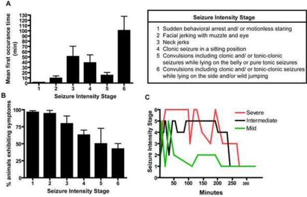
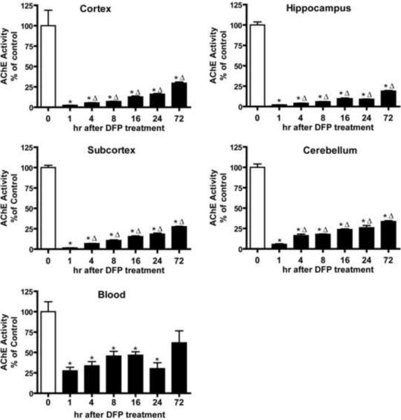
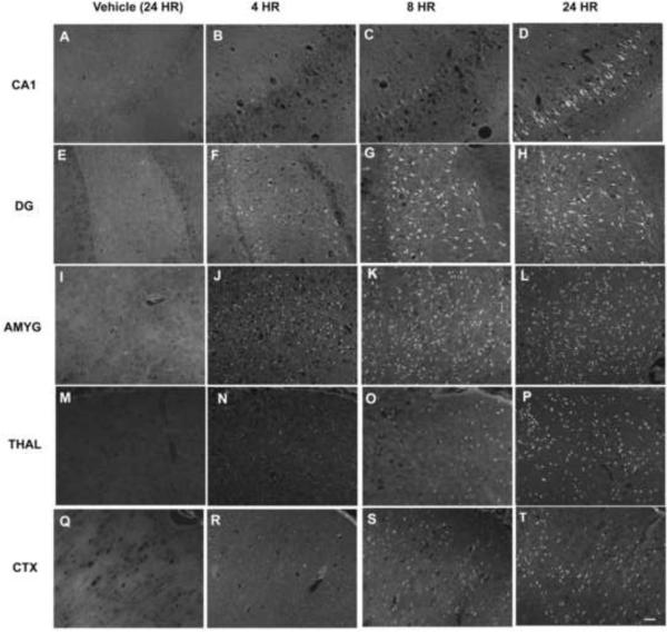
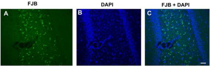
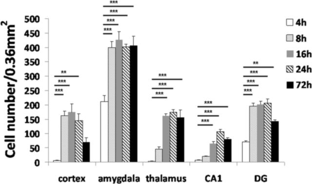
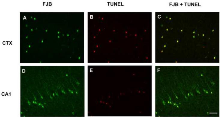
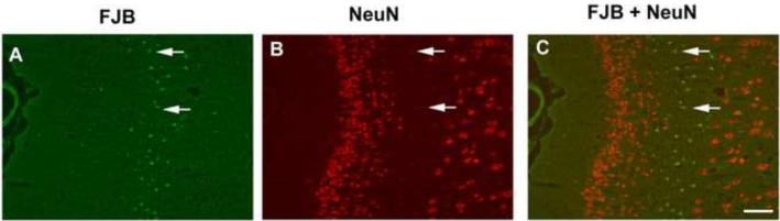
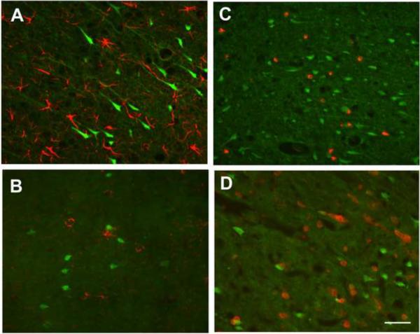
Organophosphate (OP) neurotoxins cause acute cholinergic toxicity and seizures resulting in delayed brain damage and persistent neurological symptoms. Testing novel strategies for protecting against delayed effects of acute OP intoxication has been hampered by the lack of appropriate animal models. In this study, we characterize the spatiotemporal pattern of cellular injury after acute intoxication with the OP diisopropylfluorophosphate (DFP). Adult male Sprague-Dawley rats received pyridostigmine (0.1 mg/kg, im) and atropine methylnitrate (20mg/kg, im) prior to DFP (9 mg/kg, ip) administration. All DFP-treated animals exhibited moderate to severe seizures within minutes after DFP injection but survived up to 72 h. AChE activity was significantly depressed in the cortex, hippocampus, subcortical brain tissue and cerebellum at 1h post-DFP injection and this inhibition persisted for up to 72 h. Analysis of neuronal injury by Fluoro-Jade B (FJB) labeling revealed delayed neuronal cell death in the hippocampus, cortex, amygdala and thalamus, but not the cerebellum, starting at 4h and persisting until 72 h after DFP treatment, although temporal profiles varied between brain regions. At 24h post-DFP injection, the pattern of FJB labeling corresponded to TUNEL staining in most brain regions, and FJB-positive cells displayed reduced NeuN immunoreactivity but were not immunopositive for astrocytic (GFAP), oligodendroglial (O4) or macrophage/microglial (ED1) markers, demonstrating that DFP causes a region-specific delayed neuronal injury mediated in part by apoptosis. These findings indicate the feasibility of this model for testing neuroprotective strategies, and provide insight regarding therapeutic windows for effective pharmacological intervention following acute OP intoxication.


Figures

Figure 1
Figure 1. Seizure activity in rats following acute DFP intoxication
Rats were pretreated with pyridostigmine (0.1 mg/kg, im) and atropine methyl nitrate (20 mg/kg, im) 30 or 10 min, respectively, prior to ip injection of DFP (9 mg/kg) or an equal volume (300 μl) of vehicle. Seizure behavior was monitored continuously for 5 h post-DFP injection and scored using a revised Racine's scale (Luttjohann et al., 2009) as described in the Table. Seizure behaviors were not observed in vehicle controls (data not shown). The seizure behaviors elicited by DFP in rats pretreated with pyridostigmine and atropine were variable within the treatment group (n=12) as evidenced by the mean onset time for each specific behavior following DFP injection (A) and the percentage of DFP-treated animals exhibiting any specific seizure behavior (B). (C) Representative activity profiles of rats exhibiting three different patterns of seizure behaviors following DFP injection. Data in panels A and B are expressed as X ± SE (n includes only those animals exhibiting the specific behavior).
Figure 2
Figure 2. Time course of acetylcholinesterase (AChE) inhibition in specific rat brain regions following acute DFP intoxication
Anesthetized rats were pretreated with pyridostigmine (0.1 mg/kg, im) and atropine methyl nitrate (20 mg/kg, im) 30 or 10 min, respectively, prior to injection of DFP (9 mg/kg, ip) or an equal volume (300 μl) of vehicle (water). AChE activity was determined in whole blood and specific brain regions collected immediately after euthanasia at varying times after DFP injection and raw values converted to a percentage of the mean AChE activity in corresponding tissue from vehicle controls. Data are expressed as X ± SE (n = 5 per treatment group). Statistically significant differences were determined using one way ANOVA with post hoc Tukey's test; *p<0.01 compared to vehicle control; Δp<0.01 compared to 1 h post-DFP injection.
Figure 3
Figure 3. Acute DFP intoxication elicits neuronal injury in the brain that is time and region dependent
Anesthetized rats were pretreated with pyridostigmine (0.1 mg/kg, im) and atropine methyl nitrate (20 mg/kg, im) 30 or 10 min, respectively, prior to ip injection of DFP (9 mg/kg) or an equal volume of vehicle (CTRL, panels A, E, I, M and Q). DFP-treated animals were euthanized and their brains collected for staining with Fluoro-Jade B (FJB) at 4 h (B, F, J, N and R), 8 h (C, G, K, O and S) or 24 h (D, H, L, P and T) after DFP injection. Representative photomicrographs of FJB labeling in the CA1 region of the hippocampus (CA1; A–D), hippocampal dentate gyrus (DG; E–H), amygdala (AMYG; I–L), dorsal thalamus (THAL; M–P) and somatosensory cortex (CTX; Q–T) for each treatment group. Bar = 100 μm.
Figure 4
Figure 4. Acute DFP intoxication injures a subpopulation of neurons
Representative photomicrographs of (A) Fluoro-Jade B (FJB) labeling and (B) DAPI staining in the hippocampal CA1 region of a rat 24 h post-DFP injection. All animals were anesthetized and pretreated with pyridostigmine (0.1 mg/kg, im) and atropine methyl nitrate (20 mg/kg, im) prior to DFP (9 mg/kg, ip) injection. (C) A merged image illustrates that only a subpopulation of DAPI-positive cells is labeled with FJB. Bar = 100 μm.
Figure 5
Figure 5. Acute DFP intoxication elicits neuronal injury in the brain that is time and region dependent
Anesthetized rats were pretreated with pyridostigmine (0.1 mg/kg, im) and atropine methyl nitrate (20 mg/kg, im) 30 or 10 min, respectively, prior to ip injection of DFP (9 mg/kg) or an equal volume (300 μl) of vehicle. DFP-treated rats were euthanized at varying times after DFP injection and the brains collected for staining with Fluoro-Jade B (FJB). The number of FJB-positive cells in specific brain regions at the same level as determined using a brain atlas was quantified from digital images of coronal sections (200× magnification). Control animals did not display FJB labeling (data not shown). Significant increases in FJB-positive cells were observed in multiple brain regions, although the magnitude and temporal profile varied between brain regions. Data are expressed as X ± SE (n=4 to 6 animals per treatment group). Statistically significant differences were identified using one way ANOVA with post hoc Tukey's test; **p<0.01 and ***p<0.001 compared to region-matched 4 h post-DFP injection samples.
Figure 6
Figure 6. Co-localization of Fluoro-Jade B (FJB) labeling and TUNEL labeling at 24 h post-DFP injection
Anesthetized rats were pretreated with pyridostigmine (0.1 mg/kg, im) and atropine methyl nitrate (20 mg/kg, im) 30 or 10 min, respectively, prior to ip injection of DFP (9 mg/kg). At 24 h post-DFP injection, rats were euthanized and brain sections co-labeled with FJB (green, A and D) and TUNEL (red, B and E). TUNEL labeling was examined in the amygdala, cortex, thalamus and hippocampus from three DFP-treated rats. Representative photomicrographs are shown from the cingulate cortex (CTX, panels A–C) and hippocampus CA1 (panels D–F). Merged images illustrates that most FJB-positive cells in the cingulate cortex also exhibit TUNEL labeling (C); whereas in the hippocampus CA1, a subset of cells exhibit co-labeling with FJB and TUNEL (F). Bar = 100 μm.
Figure 7
Figure 7. NeuN labeling is reduced in injured neurons at 24 h post-DFP injection
Anesthetized rats were pretreated with pyridostigmine (0.1 mg/kg, im) and atropine methyl nitrate (20 mg/kg, im) 30 or 10 min, respectively, prior to ip injection of DFP (9 mg/kg). At 24 h post-injection, rats were euthanized and brain sections labeled with Fluoro-Jade B (FJB, green, A) and immunostained for NeuN (red, B). NeuN immunoreactivity was examined in the amygdala, cortex, thalamus and hippocampus from three DFP-treated rats. Representative photomicrographs from the cingulate cortex show that in areas of neuronal injury as identified by FJB labeling (arrows in A), the relative intensity of NeuN (arrows in B) is dramatically reduced relative to large numbers of neurons with relatively high levels of NeuN immunoreactivity in uninjured brain regions (B). A merged image demonstrates FJB labeling in cells with low-NeuN expression (C; arrows). Bar = 100 μm.
Figure 8
Figure 8. Non-neuronal cells are not labeled by Fluoro-Jade B (FJB) in brains from rats acutely intoxicated with DFP
Anesthetized rats were pretreated with pyridostigmine (0.1 mg/kg, im) and atropine methyl nitrate (20 mg/kg, im) 30 or 10 min, respectively, prior to ip injection of DFP (9 mg/kg). Brain sections collected at 24 h post-injection were labeled with FJB and immunostained for the astrocyte-selective antigen GFAP (A), the oligodendrocytes antigen O4 (B), the activated macrophage antigen ED1 (C) or the microglia antigen CD11b (D). FJB labeling and immunoreactivity for these various markers were examined in the amygdala, cortex, thalamus and hippocampus from three DFP-treated rats. Representative images from the hippocampus CA1 (A), cingulate cortex (B), dentate gyrus (C) and somatosensory cortex (D) illustrate the general finding across all four brain regions that FJB-labeled cells do not co-localize with GFAP, O4, ED1 or CD11b-immunoreactivity. Bar = 100 μm.

References

    1. Allen GV, Gerami D, Esser MJ. Conditioning effects of repetitive mild neurotrauma on motor function in an animal model of focal brain injury. Neuroscience. 2000;99:93–105. - PubMed
    1. Anderson KJ, Fugaccia I, Scheff SW. Fluoro-jade B stains quiescent and reactive astrocytes in the rodent spinal cord. J Neurotrauma. 2003;20:1223–1231. - PubMed
    1. Auta J, Costa E, Davis J, Guidotti A. Imidazenil: a potent and safe protective agent against diisopropyl fluorophosphate toxicity. Neuropharmacology. 2004;46:397–403. - PubMed
    1. Baille V, Clarke PG, Brochier G, Dorandeu F, Verna JM, Four E, Lallement G, Carpentier P. Soman-induced convulsions: the neuropathology revisited. Toxicology. 2005;215:1–24. - PubMed
    1. Baille V, Dorandeu F, Carpentier P, Bizot JC, Filliat P, Four E, Denis J, Lallement G. Acute exposure to a low or mild dose of soman: biochemical, behavioral and histopathological effects. Pharmacology, biochemistry, and behavior. 2001;69:561–569. - PubMed

Publication types