. 2011 May 17;19(5):652-63.
doi: 10.1016/j.ccr.2011.04.002. Epub 2011 Apr 21.

c-Raf, but not B-Raf, is essential for development of K-Ras oncogene-driven non-small cell lung carcinoma

Affiliations

c-Raf, but not B-Raf, is essential for development of K-Ras oncogene-driven non-small cell lung carcinoma

Rafael B Blasco et al. Cancer Cell. .

Abstract

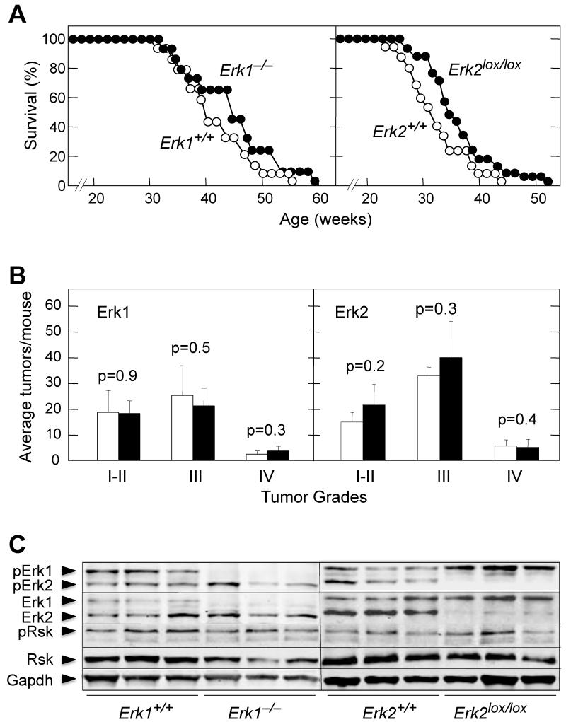
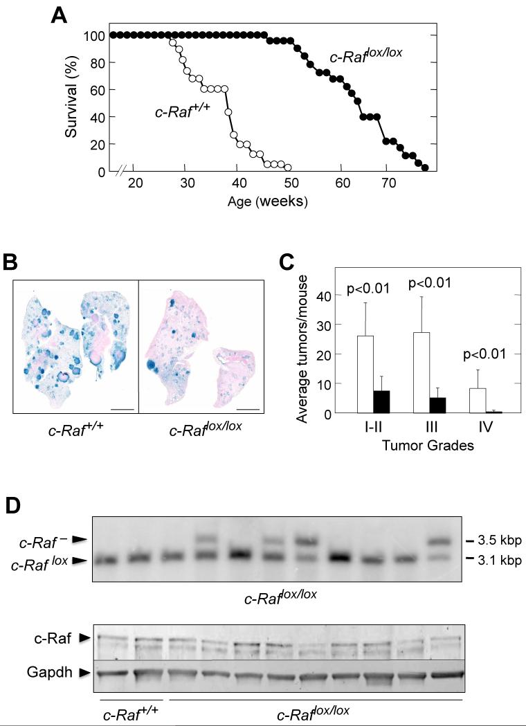
We have investigated the role of individual members of the Raf/Mek/Erk cascade in the onset of K-Ras oncogene-driven non-small cell lung carcinoma (NSCLC). Ablation of Erk1 or Erk2 in K-Ras oncogene-expressing lung cells had no significant effect due to compensatory activities. Yet, elimination of both Erk kinases completely blocked tumor development. Similar results were obtained with Mek kinases. Ablation of B-Raf had no significant effect on tumor development. However, c-Raf expression was absolutely essential for the onset of NSCLC. Interestingly, concomitant elimination of c-Raf and B-Raf in adult mice had no deleterious consequences for normal homeostasis. These results indicate that c-Raf plays a unique role in mediating K-Ras signaling and makes it a suitable target for therapeutic intervention.


Figures

Figure 1
Figure 1. K-RasG12V induces NSCLCs in the absence of Erk1 or Erk2
(A) (Left) Survival of K-Ras+/G12V;Erk1+/+ (n=17) (open circles) and K-Ras+/G12V;Erk1−/− (n=14) (solid circles) mice treated with Ad-Cre at 8 weeks of age. (Right) Survival of K-Ras+/G12V;Erk2+/+ (n=19) (open circles) and K-Ras+/G12V;Erk2lox/lox (n=32) (solid circles) mice treated with Ad-Cre at 8 weeks of age. (B) (Left) Number of tumors, classified by grade (I-IV), observed in K-Ras+/G12V;Erk1+/+ (n=5) (open bars) and K-Ras+/G12V;Erk1−/− (n=5) (solid bars) mice sacrificed 6 months after Ad-Cre treatment. (Right) Number of tumors, classified by grade (I-IV), observed in K-Ras+/G12V;Erk2+/+(n=5) (open bars) and K-Ras+/G12V;Erk2lox/lox (n=5) (solid bars) mice sacrificed 6 months after Ad-Cre treatment. Error bars indicate +/− SD of the mean. p values were calculated according to Student’s t test. (C) Western blot analysis of pErk1/2, Erk1/2, pRsk and Rsk expression in lysates prepared from individual tumors of the indicated genotype collected 8 months after Ad-Cre treatment. Gapdh is shown as a loading control. Migration of the above proteins is indicated by arrowheads. See also figure S1.
Figure 2
Figure 2. Complete ablation of Erk1/2 kinases prevents induction of NSCLCs by a resident K-RasG12V oncogene
(A) Survival of K-Ras+/G12V;Erk1+/+;Erk2+/+ (n=14) (open circles) and K-Ras+/G12V;Erk1−/−;Erk2lox/lox (n=11) (solid circles) mice treated with Ad-Cre at 8 weeks of age. (B) Whole mount X-Gal staining of lung sections collected from mice with the indicated genotypes 6 months after Ad-Cre treatment. β-Geo positive cells identified by X-Gal staining (blue color) correspond to cells expressing K-RasG12V. Scale bar, 5 mm. (C) Number of tumors, classified by grade (I-IV), observed in K-Ras+/G12V;Erk1+/+;Erk2+/+ (n=7) (open bars) and K-Ras+/G12V;Erk1−/−;Erk2lox/lox (n=10) (solid bars) mice sacrificed 6 months after Ad-Cre treatment. Error bars indicate +/− SD of the mean. p values were calculated according to Student’s t test. (D) (Top) Southern blot analysis of genomic DNA isolated from individual tumors of K-Ras+/G12V;Erk1−/−;Erk2lox/lox mice 8 months after Ad-Cre treatment. DNAs were digested with KpnI and probed with a 450 bp DNA fragment derived from the third intron just outside the floxed sequences. Migration of the unrecombined Erk2lox allele (4.3 kpb) and the ablated Erk2 allele (2.4 kbp) is indicated by arrowheads. (Bottom) Western blot analysis of Erk1 and Erk2 expression in lysates obtained from individual tumors collected 8 months after Ad-Cre treatment of K-Ras+/G12V;Erk1+/+;Erk2+/+ and K-Ras+/G12V;Erk1−/−;Erk2lox/lox mice. The presence of Erk2 in tumors of Ad-Cre treated K-Ras+/G12V;Erk1−/−;Erk2lox/lox mice, due to incomplete cleavage of the Erk2lox allele, indicates that Erk2 is essential for tumor development. Gapdh was used as loading control. Migration of the above proteins is indicated by arrowheads. (E) Survival of Erk1+/+;Erk2+/+;RERTert/ert (open circles) and Erk1−/−;Erk2lox/lox;RERTert/ert (solid circles) mice fed ad libitum a tamoxifen-containing diet to activate the knocked in CreERT2 recombinase encoded by the RERTert alleles. See also figure S2.
Figure 3
Figure 3. K-RasG12V induces NSCLCs in the absence of Mek1 or Mek2
(A) (Left) Survival of K-Ras+/G12V;Mek1+/+ (n=20) (open circles) and K-Ras+/G12V;Mek1lox/lox (n=14) (solid circles) mice treated with Ad-Cre at 8 weeks of age. (Right) Survival of K-Ras+/G12V;Mek2+/+ (n=20) (open circles) and K-Ras+/G12V;Mek2−/− (n=19) (solid circles) mice treated with Ad-Cre at 8 weeks of age. (B) (Left) Number of tumors, classified by grade (I-IV), observed in K-Ras+/G12V;Mek1+/+ (n=7) (open bars) and K-Ras+/G12V;Mek1lox/lox (n=6) (solid bars) mice sacrificed 6 months after Ad-Cre treatment. (Right) Number of tumors, classified by grade (I-IV), observed in K-Ras+/G12V;Mek2+/+ (n=5) (open bars) and K-Ras+/G12V;Mek2−/− (n=5) (solid bars) mice sacrificed 6 months after Ad-Cre treatment. Error bars indicate +/− SD of the mean. p values were calculated according to Student’s t test. (C) Western blot analysis of pMek, Mek1, Mek2, pErk1/2 and Erk1/2 expression in lysates derived from individual tumors of the indicated genotype collected 8 months after Ad-Cre treatment. Gapdh was used as loading control. Migration of the above proteins is indicated by arrowheads. See also figure S3.
Figure 4
Figure 4. Elimination of Mek1/2 kinases prevents induction of NSCLCs by an endogenous K-RasG12V oncogene
(A) Survival of K-Ras+/G12V;Mek1+/+;Mek2+/+ (n=10) (open circles) and K-Ras+/G12V;Mek1lox/lox;Mek2−/− (n=14) (solid circles) mice treated with Ad-Cre at 8 weeks of age. (B) Whole mount X-Gal staining of lung sections collected from mice with the indicated genotypes 6 months after Ad-Cre treatment. β-Geo positive cells identified by X-Gal staining (blue color) correspond to cells expressing K-RasG12V. Scale bars 5 mm. (C) Number of tumors, classified by grade (I-IV), observed in K-Ras+/G12V;Mek1+/+;Mek2+/+ (n=6) (open bars) and K-Ras+/G12V;Mek1lox/lox;Mek2−/− (n=8)(solid bars) mice. Error bars indicate +/− SD of the mean. p values were calculated according to Student’s t test. (D) (Top) Southern blot analysis of genomic DNA isolated from individual tumors of K-Ras+/G12V;Mek1lox/lox;Mek2−/− mice 8 months after Ad-Cre treatment. DNAs were digested with HindIII and probed with a 680 bp DNA fragment obtained from a region downstream from the second loxP site. The migration of the unrecombined Mek1lox allele (1.7 kbp) and the Mek1 allele (1.5 kbp) is indicated by arrowheads. (Bottom) Western blot analysis of Mek1 and Mek2 expression in lysates obtained from individual tumors collected 8 months after Ad-Cre treatment of K-Ras+/G12V;Mek1+/+;Mek2+/+ and K-Ras+/G12V;Mek1lox/lox;Mek2−/− mice. The presence of Mek1 in tumors of Ad-Cre treated K-Ras+/G12V;Mek1lox/lox;Mek2−/− mice, due to partial cleavage of the Mek1lox allele, indicates that Mek1 is essential for tumor development. Gapdh was used as loading control. Migration of the above proteins is indicated by arrowheads (E) Survival of Mek1+/+;Mek2+/+;RERTert/ert (n=6) (open circles) and Mek1lox/lox;Mek2−/−;RERTert/ert (n=6) (solid circles) mice fed ad libitum a tamoxifen-containing diet to activate the knocked in CreERT2 recombinase encoded by the RERTert alleles. See also figure S4.
Figure 5
Figure 5. B-Raf is not required for K-Ras+/G12V induced NSCLCs in mice
(A) Survival of K-Ras+/G12V;B-Raf+/+ (n=28) (open circles) and K-Ras+/G12V;B-Raflox/lox (n=25) (solid circles) mice treated with Ad-Cre at 8 weeks of age. (B) Whole mount X-Gal staining of lung sections collected from mice with the indicated genotypes 6 months after Ad-Cre treatment. β-Geo positive cells identified by X-Gal staining (blue color) correspond to cells expressing K-RasG12V. Scale bars 5 mm. (C) Number of tumors, classified by grade (I-IV), observed in K-Ras+/G12V;B-Raf+/+ (n=5) (open bars) and K-Ras+/G12V;B-Raflox/lox (n=5) (solid bars) mice. Error bars indicate +/− SD of the mean. p values were calculated according to Student’s t test. (D) Western blot analysis of B-Raf, c-Raf, A-Raf, pMek, Mek1, pErk1/2 and Erk1/2 expression in lysates derived from individual tumors of the indicated genotype collected 8 months after Ad-Cre treatment. Gapdh was used as loading control. Migration of the above proteins is indicated by arrowheads. (E) Tumors retained phosphorylated Erk expression in the absence of B-Raf. Hematoxylin & Eosin (H&E) (left) and immunohistochemical staining of consecutive paraffin fixed sections using anti-B-Raf (center) and anti-pErk (right) antibodies. Sections were obtained from lungs of Ad-Cre treated K-Ras+/G12V;B-Raf+/+ (top) and K-Ras+/G12V;B-Raflox/lox (bottom) mice. Insets show detail of larger areas. Scale bars, main field: 0.5 mm and inset: 0.02mm. See also figure S5.
Figure 6
Figure 6. c-Raf is essential for K-Ras+/G12V induced NSCLCs in mice
(A) Survival of K-Ras+/G12V;c-Raf+/+ (n=22) (open circles) and K-Ras+/G12V;c-Raflox/lox (n=23) (solid circles) mice treated with Ad-Cre at 8 weeks of age. (B) Whole mount X-Gal staining of lung sections collected from mice with the indicated genotypes 6 months after Ad-Cre treatment. β-Geo positive cells identified by X-Gal staining (blue color) correspond to cells expressing K-RasG12V. Scale bars, 5 mm. (C) Number of tumors, classified by grade (I-IV), observed in K-Ras+/G12V;c-Raf+/+ (n=8) (open bars) and K-Ras+/G12V;c-Raflox/lox (n=8) (solid bars) mice. Error bars indicate +/− SD of the mean. p values were calculated according to Student’s t test. (D) (Top) Southern blot analysis of DNA isolated from individual tumors obtained from K-Ras+/G12V;c-Raflox/lox mice infected with Ad-Cre particles at 8 weeks of age. Tumor DNAs were digested with PstI. The sizes of the diagnostic DNA fragments for c-Raflox and c-Raf alleles are indicated. (Bottom) Western blot analysis of c-Raf expression in lysates obtained from individual tumors collected 8 months after Ad-Cre treatment of K-Ras+/G12V;c-Raf+/+ and K-Ras+/G12V;c-Raflox/lox mice. The presence of c-Raf in tumors of Ad-Cre treated K-Ras+/G12V;c-Raflox/lox mice is due to incomplete cleavage of the c-Raflox allele. These results indicate that c-Raf is essential for tumor development. Gapdh was used as loading control. Migration of the above proteins is indicated by arrowheads. See also figure S6.
Figure 7
Figure 7. Adult mice tolerate well widespread ablation of B-Raf and c-Raf alleles
(A) Southern blot analysis of DNA isolated from tissues of c-Raflox/lox;RERTert/ert mice fed ad libitum with a tamoxifen diet for three months (P30 to P120). DNA was digested with PstI and probed with a 854 bp DNA fragment corresponding to sequences located in intron 4 downstream from the second loxP site. Migration of the ablated c-Raf allele and the unrecombined c-Raflox allele is indicated by arrowheads. The sizes of the diagnostic DNA fragments for these alleles are also indicated. Whereas some tissues such as testis (Te), ovaries (Ov), heart (Ht) and muscle (Mu) display partial c-Raflox cleavage (from 50 to 70%), the majority of the tissues, including lung (Lu), stomach (St), colon (Co), skin (Sk), intestine (In), liver (Li), kidney (Ki), pancreas (Pa) and spleen (Sp) showed complete or almost complete excision. Brain tissue (Br) served as negative control since tamoxifen crosses the brain-blood barrier with limited efficiency. (B) Southern blot analysis of DNA isolated from tissues of B-Raflox/lox;c-Raflox/lox;RERTert/ert mice fed ad libitum with a tamoxifen diet for three months (P30 to P120). DNA was digested with HindIII and probed with a 422 bp DNA fragment corresponding to sequences located in intron 12 upstream from the first loxP site. Migration of the ablated B-Raf null (B-Raf) and c-Raf null (c-Raf) alleles as well as of the unrecombined B-Raflox and c-Raflox alleles is indicated by arrowheads. The sizes of the diagnostic DNA fragments for these alleles are also indicated. (Top) B-Raflox alleles are almost completely recombined in lung (Lu), stomach (St), colon (Co), kidney (Ki), pancreas (Pa), spleen (Sp) and thymus (Th) and only partially cleaved in testis (Te), ovaries (Ov) and heart (Ht). (Bottom) c-Raflox alleles were partially excised in testis (Te), ovaries (Ov), heart (Ht), lung (Lu), stomach (St) and colon (Co) and completely recombined in kidney (Ki), pancreas (Pa) and spleen (Sp) and thymus (Th). Brain tissue (Br) served as negative control. See also figure S7.
Figure 8
Figure 8. B-Raf and c-Raf are not essential for proliferation and immortalization of primary MEFs driven by wild type or oncogenic K-Ras signaling
(A) Western blot analysis of B-Raf and c-Raf protein expression in primary MEFs derived from (top) three independent K-Ras+/G12V;RERTert/ert, K-Ras+/G12V;B-Raflox/lox,RERTert/ert, K-Ras+/G12V;c-Raflox/lox;RERTert/ert and K-Ras+/G12V;B-Raflox/lox;c-Raflox/lox;RERTert/ert embryos and (bottom) three independent K-Ras+/+;RERTert/ert, K-Ras+/+;B-Raflox/lox;RERTert/ert, K-Ras+/+;c-Raflox/lox;RERTert/ert and K-Ras+/+;B-Raflox/lox;c-Raflox/lox;RERTert/ert embryos. These MEFs were incubated in DMEM media supplemented with 10% FBS in the presence of 4OHT for 5 days to activate expression of the resident K-RasG12V oncogene and to eliminate the c-Raflox and/or B-Raflox conditional alleles. Gapdh served as loading control. Migration of the corresponding proteins is indicated by arrowheads. (B) Growth curves of the primary MEFs (n=3) described above. MEFs were grown in DMEM media supplemented with 10% FBS and 600 nM 4OHT during 5 days before seeding. Results are shown in arbitrary absorbance units. Left panel: K-Ras+/G12V;RERTert/ert (solid circles), K-Ras+/G12V;B-RafΔ/Δ;RERTert/ert (open squares), K-Ras+/G12V;c-RafΔ/Δ;RERTert/ert (open circles), K-Ras+/G12V;B-RafΔ/Δ;c-RafΔ/Δ;RERTert/ert (open triangles). Right panel: K-Ras+/+;RERTert/ert (solid circles), K-Ras+/+;B-RafΔ/Δ;RERTert/ert (open squares), K-Ras+/+;c-RafΔ/Δ;RERTert/ert (open circles), K-Ras+/+;B-RafΔ/Δ;c-RafΔ/Δ;RERTert/ert (open triangles). Error bars indicate +/− SD of the mean. (C) (Left) Western blot analysis of B-Raf and c-Raf protein expression in primary MEFs derived from five independent K-Ras+/G12V;B-Raflox/lox;RERTert/ert, K-Ras+/G12V;c-Raflox/lox;RERTert/ert and K-Ras+/G12V;B-Raflox/lox;c-Raflox/lox;RERTert/ert embryos. Samples from two K-Ras+/G12V;RERTert/ert embryos are also shown. MEFs were incubated in DMEM media supplemented with 2% FBS in the presence of 4OHT for 5 days to activate expression of the resident K-RasG12V oncogene and to eliminate the c-Raflox and/or B-Raflox conditional alleles. Gapdh served as loading control. Migration of the corresponding proteins is indicated by arrowheads. (Right) Growth curves of the primary MEFs (n=5) described above. MEFs were grown in DMEM media supplemented with 2% FBS and 600 nM 4OHT. K-Ras+/G12V;RERTert/ert (solid circles), K-Ras+/G12V;B-RafΔ/Δ;RERTert/ert (open squares), K-Ras+/G12V;c-RafΔ/Δ;RERTert/ert (open circles), K-Ras+/G12V;B-RafΔ/Δ;c-RafΔ/Δ;RERTert/ert(open triangles). Error bars indicate +/− SD of the mean. (D) Immortalization of the primary MEFs described in (B) following a 3T3 protocol (n=3). Cells were cultivated in DMEM media supplemented with 10% FBS and 600 nM 4OHT. Symbols are those described in (B). Error bars indicate +/− SD of the mean. See also figure S8.

References

    1. Barbie DA, Tamayo P, Boehm JS, Kim SY, Moody SE, Dunn IF, Schinzel AC, Sandy P, Meylan E, Scholl C, et al. Systematic RNA interference reveals that oncogenic KRAS-driven cancers require TBK1. Nature. 2009;462:108–112. - PMC - PubMed
    1. Belanger LF, Roy S, Tremblay M, Brott B, Steff AM, Mourad W, Hugo P, Erikson R, Charron J. Mek2 is dispensable for mouse growth and development. Mol Cell Biol. 2003;23:4778–4787. - PMC - PubMed
    1. Bissonauth V, Roy S, Gravel M, Guillemette S, Charron J. Requirement for Mak2k1 (Mek1) in extra-embryonic ectoderm placentogenesis. Development. 2006;133:3429–3440. - PubMed
    1. Bollag G, Hirth P, Tsai J, Zhang J, Ibrahim PN, Cho H, Spevak W, Zhang C, Zhang Y, Habets G, et al. Clinical efficacy of a RAF inhibitor needs broad target blockade in BRAF-mutant melanoma. Nature. 2010;467:596–599. - PMC - PubMed
    1. Camonis JH, White MA. Ral GTPases: corrupting the exocyst in cancer cells. Trends Cell Biol. 2005;15:327–332. - PubMed

Publication types

MeSH terms