

Human proteins with affinity for dermatan sulfate have the propensity to become autoantigens
- PMID: 21514432
- PMCID: PMC3081203
- DOI: 10.1016/j.ajpath.2011.01.031
Human proteins with affinity for dermatan sulfate have the propensity to become autoantigens
Abstract
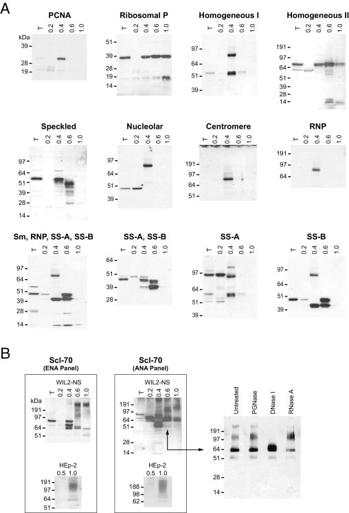
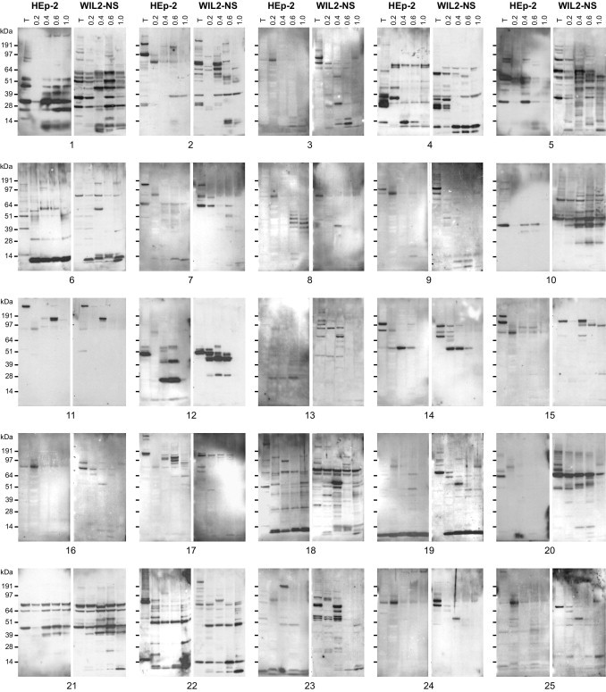
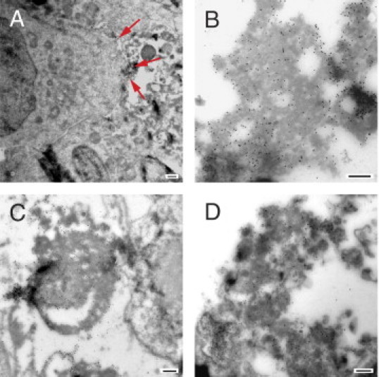
The mystery of why and how a small, seemingly disparate subset of all self molecules become functional autoantigens holds a key to understanding autoimmune diseases. Here and in a companion article in this issue, we show that affinity of self molecules to the glycosaminoglycan dermatan sulfate (DS) is a common property of autoantigens and leads to a specific autoreactive B-1a cell response. Autoimmune ANA/ENA reference sera react preferentially with DS affinity-fractionated cellular proteins. Studying patients with autoimmune diseases, we discovered patient-specific complex autoantigen patterns that are far richer and more diverse than previously thought, indicating significant pathological heterogeneity even within traditionally defined clinical entities, such as systemic lupus erythematosus. By shotgun sequencing of DS affinity-enriched proteomes extracted from cell lines, we identified approximately 200 autoantigens, both novel and previously linked to autoimmunity, including several well-known families of autoantigens related to the nucleosome, ribonucleoproteins, the cytoskeleton, and heat shock proteins. Using electron microscopy, we recognized direct interaction with dead cells as an origin of autoantigenic association of DS with self molecules. DS affinity may be a unifying property of the human autoantigen-ome (ie, totality of self molecules that can serve as functional autoantingens) and thus provides a promising tool for discovery of autoantigens, molecular diagnosis of autoimmune diseases, and development of cause-specific therapies.
Copyright © 2011 American Society for Investigative Pathology. Published by Elsevier Inc. All rights reserved.
Figures
References
-
- Plotz P.H. The autoantibody repertoire: searching for order. Nat Rev Immunol. 2003;3:73–78. - PubMed
-
- Amital H., Shoenfeld Y. Natural autoantibodies, heralding, protecting and inducing autoimmunity. In: Shoenfeld Y., Gershwin M.E., Meroni P.L., editors. Autoantibodies. ed 2. Elsevier; Amsterdam: 2007. pp. 7–12.
-
- Gilbert D., Brard F., Jovelin F., Tron F. Do naturally occurring autoantibodies participate in the constitution of the pathological B-cell repertoire in systemic lupus erythematosus? J Autoimmun. 1996;9:247–257. - PubMed
-
- Cohen P.L., Maldonado M.A. Animal models for SLE. Curr Protoc Immunol. 2003 - PubMed
-
Chapter 15:Unit 15.20
Publication types
MeSH terms
Substances
LinkOut - more resources
Full Text Sources
