. 2011 Apr 29;145(3):357-70.
doi: 10.1016/j.cell.2011.04.002.

NEMO/NLK phosphorylates PERIOD to initiate a time-delay phosphorylation circuit that sets circadian clock speed

Affiliations

NEMO/NLK phosphorylates PERIOD to initiate a time-delay phosphorylation circuit that sets circadian clock speed

Joanna C Chiu et al. Cell. .

Erratum in

  • Cell. 2011 May13;145(4):635

Abstract

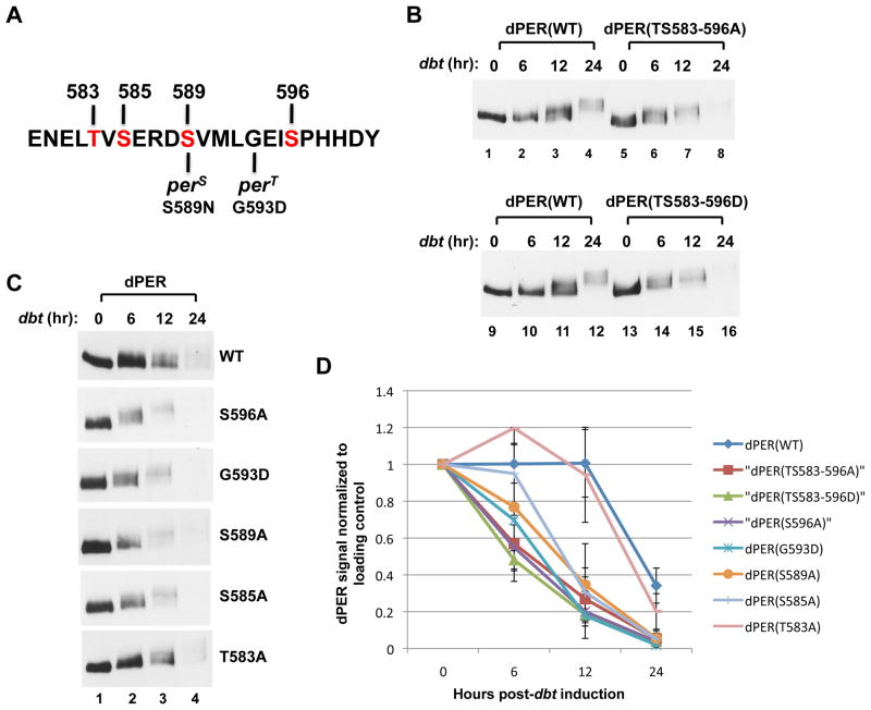
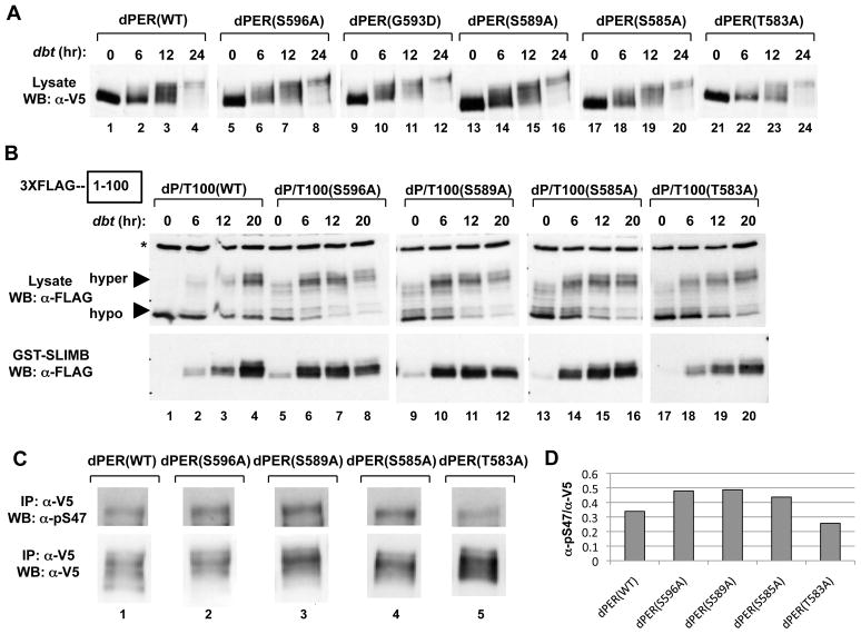
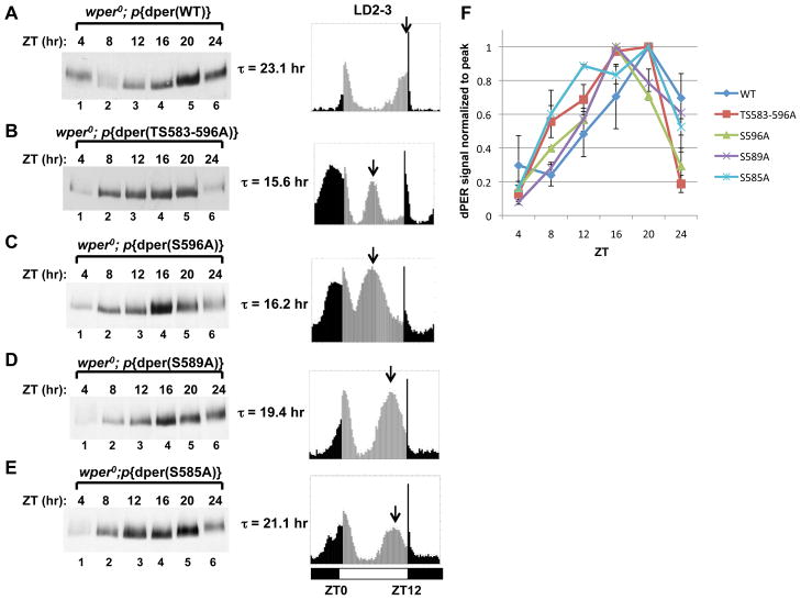
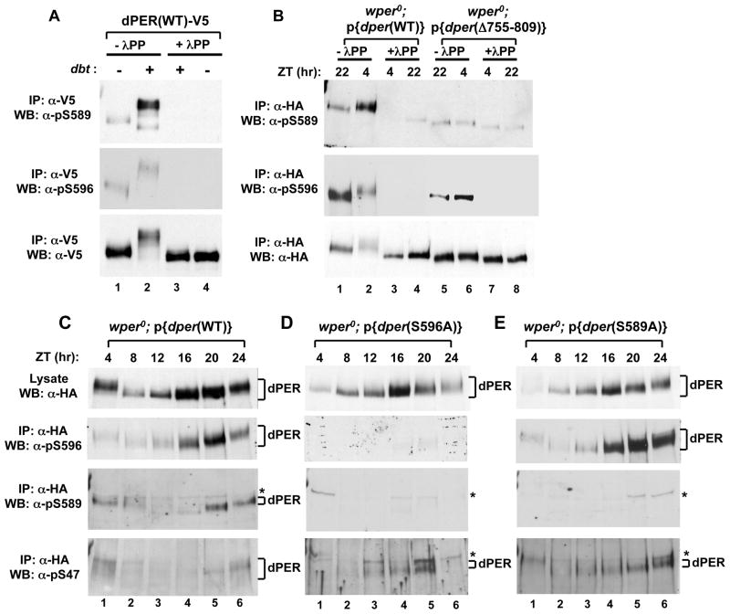
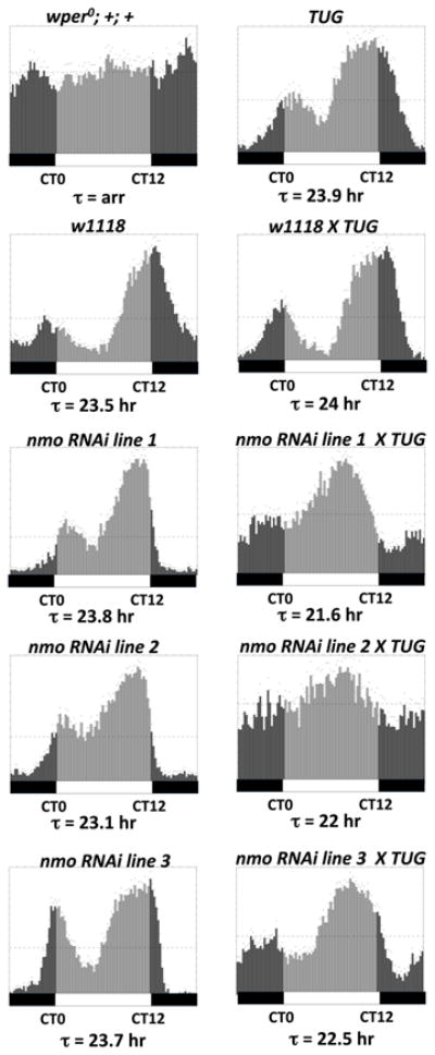
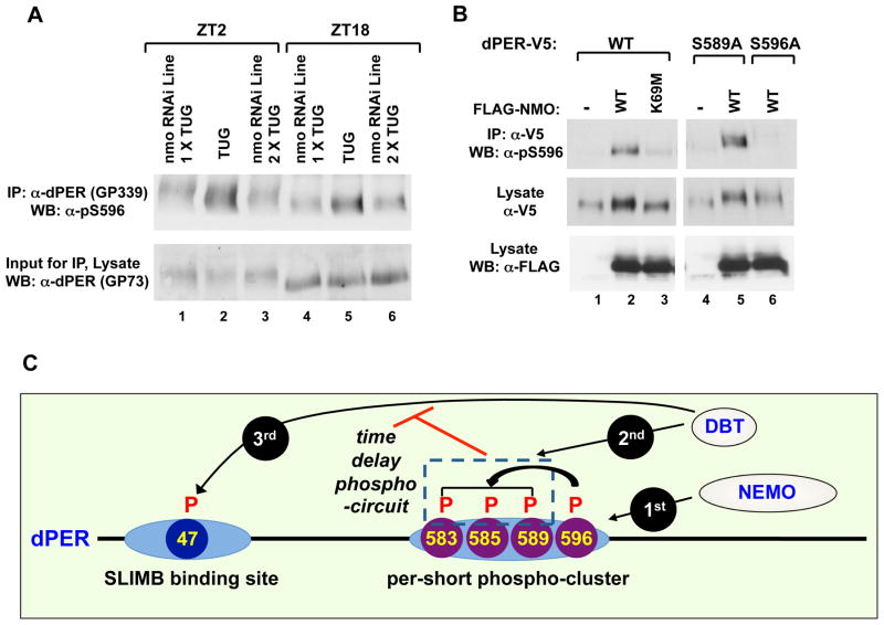
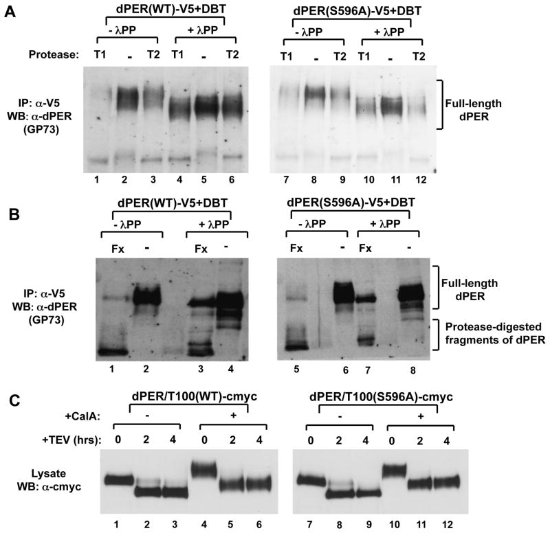
The speed of circadian clocks in animals is tightly linked to complex phosphorylation programs that drive daily cycles in the levels of PERIOD (PER) proteins. Using Drosophila, we identify a time-delay circuit based on hierarchical phosphorylation that controls the daily downswing in PER abundance. Phosphorylation by the NEMO/NLK kinase at the "per-short" domain on PER stimulates phosphorylation by DOUBLETIME (DBT/CK1δ/ɛ) at several nearby sites. This multisite phosphorylation operates in a spatially oriented and graded manner to delay progressive phosphorylation by DBT at other more distal sites on PER, including those required for recognition by the F box protein SLIMB/β-TrCP and proteasomal degradation. Highly phosphorylated PER has a more open structure, suggesting that progressive increases in global phosphorylation contribute to the timing mechanism by slowly increasing PER susceptibility to degradation. Our findings identify NEMO as a clock kinase and demonstrate that long-range interactions between functionally distinct phospho-clusters collaborate to set clock speed.


Conflict of interest statement

The authors declare no conflicts of interest.

Figures

Figure 1
Figure 1. Differential effects of phospho-site mutations in the perS phospho-cluster on DBT-dependent phosphorylation kinetics and degradation rate of dPER in cultured S2 cells
(A) Sequence of dPER from amino acid 579 to 601, encompassing the original “per-short” region (aa 585 to 601), highlighting the phosphorylation site mutations generated in this study (labeled in red) as well as locations of the perS and perT mutations. (B, C) S2 cells were co-transfected with wild type (WT) or mutant variants of pAc-3XFLAG-His-dper-6Xcmyc (simplified as dper) and pMT-dbt(WT)-V5-His (simplified as dbt, left of panels), and collected at the indicated times (hr) post-dbt induction. The dPER variants (top of panels) were detected by immunoblotting in the presence of α-c-myc antibodies. (D) dPER staining intensity values normalized to Hsp70 loading control. At least two independent experiments were used to generate the average for each time point. Error bars represent SEM. See Fig. S1 for supporting data.
Figure 2
Figure 2. Differential effects of phospho-site mutants in the perS phospho-cluster on dPER/SLIMB interactions and S47 phosphorylation
(A) S2 cells were co-transfected with dbt and dper variants and collected at the indicated times (hr) post-dbt induction (top of panels). The culture media contained MG132 to inhibit degradation of dPER. (B) S2 cells were co-transfected with WT or mutant variants of pAc-3XFLAG-His-dper/Tev100-6Xc-myc and pMT-dbt-V5-His, treated with MG132, and collected at the indicated times (hr) post-dbt induction (top of panels). Extracts were subjected to TEV protease cleavage. Equal amounts of cleaved protein were incubated with glutathione resins bound with GST-SLIMB. The relative amounts of the dPER(aa1–100) fragment in the starting material (lysate; top panels) and that bound to the resins (GST-SLIMB; bottom panels) were visualized by immunoblotting in the presence of αFLAG antibodies. Non-specific bands in the lysate (top panels) are marked by an asterisk (*, left of panel). (C) S2 cells co-expressing pAc-dper-V5-His (WT and mutants) and pMT-dbt-V5-His were incubated with MG132 and harvested at 6 hours post-dbt induction. Extracts were immunoprecipitated with α-V5 antibodies and dPER isoforms phosphorylated at Ser47 detected using α-pS47 antibodies (top), while total dPER protein was visualized using α-V5 antibodies (bottom). (D) dPER total protein and pS47 signal intensity in panel (C) were quantified, and the ratio of dPER pS47 over total dPER proteins were presented in histogram format. See Fig. S2 for supporting data.
Figure 3
Figure 3. Spatially oriented and graded responses of per-short phospho-cluster site mutants on clock speed
(A to E, left panels) Flies bearing different dper transgenes (indicated at top of panels) in a wper0 genetic background were collected at the indicated times (top of panels) and head extracts analyzed for dPER by immunoblotting in the presence of αHA. (A to E, right panels) The corresponding locomotor activity profiles of the transgenic flies in LD are shown. The second and third days worth of activity data during LD were averaged, generating the 24-hr profiles shown in the panels. The free-running periods (τ) are also shown. Arrow represents the timing of the clock-controlled evening peak. Vertical bars represent the activity recorded in 15-minute bins during times when the lights were on (grey bars) or off (black bars). ZT0 is lights-on, whereas ZT12 is lights-off. Horizontal bars at bottom; open, lights-on; closed, lights-off. (F) dPER signal intensity (Y-axis) was normalized by setting the peak intensity value for each dPER variant to a value of 1. Two independent experiments were used to generate the average and error bars represent SEM. See Table S1 for supporting data.
Figure 4
Figure 4. Phospho-specific antibodies reveal that DBT phosphorylates S589 but not S596, and that phosphorylation at S596 stimulates phosphorylation at S589 and S585, which delays phosphorylation at S47
(A) Extracts were prepared from S2 cells co-transfected with a wild type (WT) version of dper (pAc-dper-V5-His) and pMT-dbt-V5-His (+) or an empty pMT plasmid, pMT-V5-His (−). Cells were treated with MG132 and cycloheximide 16 hours post-dbt induction and harvested 4 hrs later. dPER was immunoprecipitated (IP) with α-V5 beads and split into equal parts that were either treated in the absence (-λPP) or presence (+λPP) of λ-phosphatase. Recovered immune complexes were probed by western blotting (WB) in the presence of the indicated antibody (left of panel). (B) Head extracts were prepared from wper0;p{dper(WT)} or wper0;p{der(Δaa755-809)} flies collected at the indicated times (ZT; top of panels). dPER-HAHis-containing immune complexes were recovered using α-HA beads and split into two equal parts that were treated in the absence (-λPP) or presence (+λPP) of λ-phosphatase. Recovered immune complexes were probed by western blotting (WB) in the presence of the indicated antibody (left of panel). (C, D, E) Head extracts were prepared from wper0;p{dper(WT)}, wper0;p{dper(S589A)}, and wper0;p{dper(S596A)} flies collected at the indicated times (ZT). dPER-HAHis-containing immune complexes were recovered using α-HA beads, followed by western blotting (WB) in the presence of the indicated antibody (left of panel). Positions of non-specific signals from α-pS589 and α-pS47 antibodies are indicated by asterisks (*; right of panels). See Figs. S4 and S5 for supporting data.
Figure 5
Figure 5. RNAi knockdown of nmo in pacemaker cells speeds up the pace of the clock
Shown are group averages of the 24-hr activity profiles (average of first three days during constant dark conditions) for males of the different genotypes. The free-running periods (τ) are also shown. Vertical bars represent the activity recorded in 15-minute bins during the subjective day (light grey bars; CT0-12) or subjective night (dark grey bars; CT12-24). Horizontal bars at bottom; open, subjective day; closed, subjective night. Targeted expression of nmo RNAi in tim-expressing clock neurons was achieved using the UAS/GAL4 system. The GAL4 driver line used here is w; UAS-DICER2; tim- -UAS-GAL4 (TUG). Three independent responder RNAi lines (line 1= v101545, line 2 = v104885, line 3 = v3002) were used and they were all in w1118 genetic background. Appropriate parental and genetic background (w1118) controls were included in the experiment. wper0; +; + represents the control for arrhythmicity (arr).
Figure 6
Figure 6. Nmo is a clock kinase that phosphorylates S596 on dPER
(A) Targeted expression of nmo RNAi in tim-expressing clock neurons was achieved by crossing w; UAS-DICER2; tim-UAS-GAL4 (TUG) males with females from two independent responder RNAi lines [v101545 (line 1) and v104885 (line 2)] that are in a w1118 genetic background. Flies were collected at ZT 2 or 18, and head extracts prepared. dPER-containing immune complexes were recovered using α-dPER antibodies (GP339), followed by western blotting (WB) with α-pS596 (top). The relative input of dPER for the IP was assayed by western blotting (WB) with α-dPER (GP73; bottom). (B) S2 cells were transfected with pAc-dper-V5-His (WT, S589A, or S596A mutant) in the presence of an empty plasmid (pAc-3XFLAG-His) or nmo-expressing plasmid [pAc-3XFLAG-His-nmo (WT or K69M kinase-dead mutant)]. dPER-V5-containing immune complexes were recovered using α-V5 beads, followed by western blotting (WB) in the presence of the indicated antibody (left of panel) to detect dPER isoforms phosphorylated at S596 (top panel), total dPER proteins (middle) and NMO (bottom). (C) Model showing how the per-short phospho-cluster regulates the pace of the clock. NMO phosphorylates S596 on dPER, which stimulates DBT-dependent phosphorylation of S589, S585 and (perhaps) T583. Phosphorylation in the per-short phospho-cluster somehow delays phosphorylation at other DBT-dependent sites on dPER, including S47, thus delaying the dPER/SLIMB interaction and the daily downswing in dPER levels. See Figs S4 to S6 for supporting data.
Figure 7
Figure 7. Limited proteolysis suggests highly phosphorylated dPER has a more open conformation compared to hypo-phosphorylated isoforms
(A, B) Extracts were prepared from S2 cells co-transfected with WT of S596A variants of pAc-dper-V5-His and pMT-dbt that were harvested 20 hr after dbt induction. dPER protein was immunoprecipitated (IP) with α-V5 beads and subsequently treated in the absence (-λPP) or presence (+λPP) of λ-phosphatase. Immunoblotting with α-dPER antibodies was used to detect dPER proteins after limited proteolysis using (A) Trypsin (T1 and T2) or (B) Factor Xa (Fx). (C) Extracts were prepared from S2 cells transfected with WT or S596A variants of pAc-dper/Tev100-V5-His. Hyperphosphorylated dPER was generated by addition of 30nM of Calyculin A (Calbiochem) 2 hours before cell harvest. 50uM MG132 (Sigma) and 10ug/ml cycloheximide (Sigma) were also added to maximize the amount of hyperphosphorylated dPER and to prevent degradation. Cells were harvested 48 hrs after transfection. Immunoblotting with α-c-myc antibodies was used to detect dPER (full length and aa101-1224) proteins after limited proteolysis using AcTEV (Invitrogen).

References

    1. Allada R, Chung BY. Circadian organization of behavior and physiology in Drosophila. Annu Rev Physiol. 2010;72:605–624. - PMC - PubMed
    1. Bae K, Edery I. Regulating a circadian clock's period, phase and amplitude by phosphorylation: insights from Drosophila. J Biochem. 2006;140:609–617. - PubMed
    1. Baker CL, Kettenbach AN, Loros JJ, Gerber SA, Dunlap JC. Quantitative proteomics reveals a dynamic interactome and phase-specific phosphorylation in the Neurospora circadian clock. Mol Cell. 2009;34:354–363. - PMC - PubMed
    1. Baylies MK, Bargiello TA, Jackson FR, Young MW. Changes in abundance or structure of the per gene product can alter periodicity of the Drosophila clock. Nature. 1987;326:390–392. - PubMed
    1. Baylies MK, Vosshall LB, Sehgal A, Young MW. New short period mutations of the Drosophila clock gene per. Neuron. 1992;9:575–581. - PubMed

Publication types

MeSH terms

LinkOut - more resources