. 2011 Jun 15;354(2):253-66.
doi: 10.1016/j.ydbio.2011.04.004. Epub 2011 Apr 16.

Connexin37 and Connexin43 deficiencies in mice disrupt lymphatic valve development and result in lymphatic disorders including lymphedema and chylothorax

Affiliations

Connexin37 and Connexin43 deficiencies in mice disrupt lymphatic valve development and result in lymphatic disorders including lymphedema and chylothorax

John D Kanady et al. Dev Biol. .

Abstract

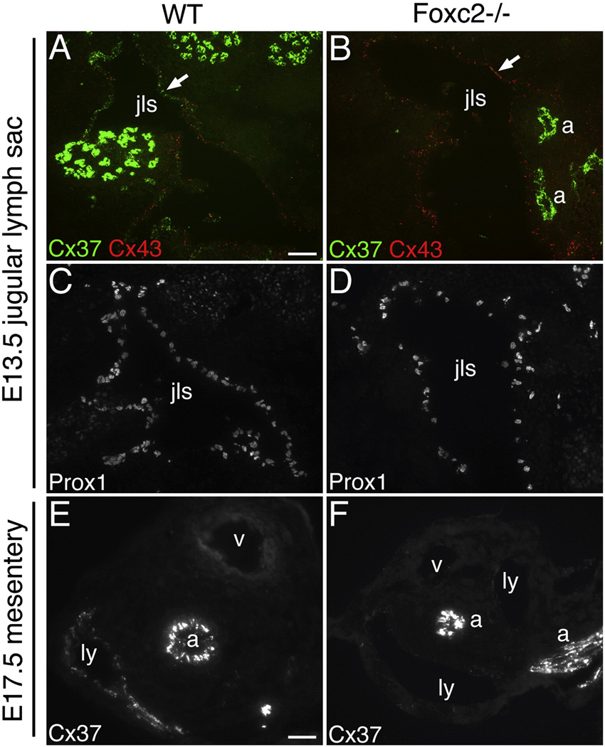
Intraluminal valves are required for the proper function of lymphatic collecting vessels and large lymphatic trunks like the thoracic duct. Despite recent progress in the study of lymphvasculogenesis and lymphangiogenesis, the molecular mechanisms controlling the morphogenesis of lymphatic valves remain poorly understood. Here, we report that gap junction proteins, or connexins (Cxs), are required for lymphatic valvulogenesis. Cx37 and Cx43 are expressed early in mouse lymphatic development in the jugular lymph sacs, and later in development these Cxs become enriched and differentially expressed by lymphatic endothelial cells on the upstream and downstream sides of the valves. Specific deficiencies of Cx37 and Cx43 alone or in combination result in defective valve formation in lymphatic collecting vessels, lymphedema, and chylothorax. We also show that Cx37 regulates jugular lymph sac size and that both Cx37 and Cx43 are required for normal thoracic duct development, including valve formation. Another Cx family member, Cx47, whose human analog is mutated in some families with lymphedema, is also highly enriched in a subset of endothelial cells in lymphatic valves. Mechanistically, we present data from Foxc2-/- embryos suggesting that Cx37 may be a target of regulation by Foxc2, a transcription factor that is mutated in human lymphedema-distichiasis syndrome. These results show that at least three Cxs are expressed in the developing lymphatic vasculature and, when defective, are associated with clinically manifest lymphatic disorders in mice and man.


Figures

Figure 1
Figure 1. Expression of Cx37, Cx43, and Cx47 in developing lymphatic vessels of the wild-type mouse embryo
(A–E) Immunolabeling of jugular lymph sac (jls) of E13.5 WT mouse embryos. Cx37 (green) and Cx43 (red) are differentially expressed in distinct domains in the JLS. The Cx37 antibody crossreacts with some muscle fiber types (upper right and lower right in panel A). (C) A higher magnification view of the JLS in (A). (D) Cx37 expression in this area is associated with a sprouting region of the JLS. (B,E) Prox1 labeling of sections adjacent to those in (A,D), respectively. (F–K) Immunolabeling of dermis and subdermis of E16.5 WT embryos. Cx37 and Cx43 (both green) is present in dermal lymphatics (ly) (F,H) as well as in deeper subcutaneous lymphatics (G,I). Cx37 is also highly expressed in arteries (a). Cx47 is weakly detected (arrows) in some E16.5 dermal (J) and subcutaneous lymphatics (K). (L–Q) Immunolabeling of thoracic duct (td) cross sections of E18.5 WT embryos. Cx37 and Cx43 are both present in the E18.5 TD (the TD lumen is collapsed in these sections) and there is significant colocalization of the Cx immunosignals (N). (O) Cx47 is weakly detected (arrow) in the E18.5 TD. (P,Q) VEGFR-3 and Prox1 labeling of adjacent sections to those in (L–O). Scale bars: (A,B) 50 µm; (C) 50 µm; (D, E) 50 µm; (F–K) 50 µm; (L–Q) 25 µm.
Figure 2
Figure 2. Cx37, Cx43, and Cx47 become progressively enriched at mesenteric lymphatic valves and are differentially expressed in mesenteric valve leaflets
(A–G) Immunolabeling of mesenteric collecting lymphatics (ly) of E18.5 WT embryos. Whole-mount immunolabeling shows Cx37 and Cx43 labeling throughout the Ly endothelium, including at valves (arrows). (B,D) CD31 labeling of the same sections shown in (A,C) respectively. Arteries are labeled (a). (E,F) Higher magnification of Cx37 and Cx43 labeling shows that the Cx immunosignals outline individual endothelial cells. (G) Cx37 (red) and Cx43 (green) colocalize in this transverse section through a mesenteric Ly vessel (non-valve region). The far-right panel shows Prox1 labeling of an adjacent section. (H,I) At P7, Cx37 is enriched at mesenteric Ly valves (arrows) whereas Cx43 is more uniformly expressed in the Ly endothelium. (J,K) At postnatal 4-weeks, Cx37 (red) and Cx43 (green) is highly enriched at mesenteric Ly valves and is differentially localized in the valve leaflets. A flat, en face view of a valve leaflet is shown in (J) and a transverse section of a leaflet is shown in (K). Cx43 is enriched in the upstream side of the leaflet (u) and Cx37 is enriched in the downstream side (d). (L) Labeling of an oblique section through the wall of a 4-week postnatal mesenteric Ly shows that Cx37 and Cx43 colocalize in non-valve Ly endothelium. (M–P) Cx47 expression in mesenteric lymphatics at different stages of development. (M) At E18.5, Cx47 is found in a subset of LECs in the mesenteric lymphatics. (N) At P3, Cx47 is found associated with valve leaflets (arrowheads). (O) At 4 weeks, Cx47 is highly restricted to a subset of cells within valve leaflets (arrowheads). In valve LECs expressing Cx47, Cx47 and Cx43 colocalize (arrowheads in panel P). In (M–O) VEGFR-3 labeling identifies the lymphatics. Scale bars: (A–D) 50 µm; (E, F) 10 µm; (G) 10 µm; (H, I) 50 µm; (J, L) 10 µm; (K) 5 µm; (M–O) 10 µm; (P) 10 µm.
Figure 3
Figure 3. Cx37, Cx43, and Cx47 are highly enriched in lymphatic valves in the adult mouse and are differentially expressed in upstream and downstream sides of thoracic duct valves
(A–H) Immunolabeling of thoracic duct (td) transverse sections from adult WT mice for Cx37 and Cx43. Cx37 and Cx43 are both detected in the endothelium of the TD general wall (non-valve region) and colocalize there (D). (C) Cx37 expression is comparatively much lower in TD than in arteries (a). (E,F) Cx37 and Cx43 are highly enriched in valves of the TD. (G) Epifluorescent imaging of a flat, en face TD valve section colabeled for Cx37 (green) and Cx43 (red) shows that the two Cxs are differentially localized. (H) Confocal z-stack imaging of another flat, en face TD valve section colabeled for Cx37 and Cx43 shows that Cx43 is enriched in the upstream side of the leaflet and Cx37 is enriched in the downstream side. In the center is the x-y projection through the z-stack. At the top is the x-z projection and at the right is the y-z projection, both showing separation of the Cx37 and Cx43 signals. (I,J) Cx47 is found exclusively in a subset of cells of the valve in the TD (a flat, en face portion of the valve is shown), where it colocalizes with Cx43 (arrowheads in J). (K–N) Immunolabeling of Cx37 and Cx43 in the ear of adult mice. Cx37 and Cx43 (both green) are detected in valved collecting lymphatics of the ear (arrows in K and L) but not in the Ly capillaries highly expressing LYVE-1 (red) (arrows in M and N). In (I,K,L) VEGFR-3 labeling highlights the lymphatics. Scale bars: (A, B) 10 µm; (C) 10 µm; (D) 10 µm; (E, F) 20 µm; (G–I) 10 µm; (J) 5 µm; (K, L) 10 µm; (M, N) 10 µm.
Figure 4
Figure 4. Cx37−/− and Cx37−/−Cx43−/− embryos have enlarged jugular lymph sacs at E13.5
(A) H&E stained transverse sections of E13.5 mouse embryos. Sections were taken just above the point where the cervical nerve crosses the JLS. The paired right and left JLS lumen is outlined in red for each genotype.. (B) Cross-sectional area measures of the JLS for the various genotypes show that the JLS is significantly enlarged in Cx37−/− and Cx37−/−Cx43−/− embryos compared to WT (asterisk indicates p < 0.05). Error bars indicate s.e.m. (C) 3D volume reconstruction of JLS (green) and cardinal vein (blue) from serially sectioned WT and Cx37−/−Cx43−/− E13.5 embryos. The volume of the JLS in the Cx37−/−Cx43−/− embryo was much larger (~6-fold) than in the WT control. Axes: z (blue), dorsal; y (green), caudal; x (red), lateral. Scale bar: (A) 100 µm.
Figure 5
Figure 5. Cx37−/−Cx43−/− embryos exhibit lymphedema and dilated superficial lymphatics
(A–C) Upper portion of embryos at E18.5. Cx37−/−Cx43−/− embryos exhibit edema whereas Cx37−/− and Cx43−/− embryos do not. (D,E) H&E stained transverse sections of E16.5 embryos show evidence of massive edema (thickening of the subcutaneous tissue is marked by a double arrow) in the Cx37−/−Cx43−/− embryo (D) but not in the WT control (E). (F,G) Transverse sections of E16.5 embryos labeled for LYVE-1 (red) and CD31 (green). The LYVE-1 labeling reveals widely dilated superficial lymphatics (arrows) in the Cx37−/−Cx43−/− embryo (F) but not in the WT control (G). Scale bars: (D,E) 400 µm; (F, G) 100 µm.
Figure 6
Figure 6. Central lymphatic patterning is abnormal in Cx43−/− and Cx37−/−Cx43−/− embryos at E18.5
Prox1 whole-mount immunostaining of E18.5 thoracic duct (td) (A–D), E18.5 intercostal Ly trunks (E, F), and E18.5 diaphragm muscle (G, H). Cx43−/− and Cx37−/−Cx43−/− TDs have extremely erratic caliber, blind-ended outcroppings, and bifurcated segments compared with WT and Cx37−/− TDs. The intercostal trunk Lys (arrows) in a Cx37−/−Cx43−/− embryo (F) are sac-like compared to the WT control (E). The Ly network (arrows) on the thoracic surface of the diaphragm muscle is diminished in the Cx37−/−Cx43−/−embryo compared to the WT control. Scale bars: (A–D) 200 µm; (E, F) 200 µm; (G, H) 200 µm.
Figure 7
Figure 7. Cx37+/−Cx43−/− and Cx37−/−Cx43−/− embryos display blood-filled lymphatics
(A) Bloody discoloration in superficial skin lymphatics of a Cx37+/−Cx43−/− E18.5 embryo. (B) Higher magnification view of blood in superficial skin lymphatics of a Cx37+/−Cx43−/− E18.5 embryo. (C) Whole-mount skin Prox1 (green) immunostaining of a Cx37−/−Cx43−/− embryo confirms the Ly identity of the bloody vessels. Blood (red) was detected by autofluorescence with UV excitation. (D) Bloody discoloration in the TD (arrow) and intercostal Ly trunks (arrowhead) of a Cx37−/−Cx43−/− E18.5 embryo. Note: the specimen was photographed after whole-mount immunostaining for Prox1 (faint spots) and blood that was in the aorta has been washed away during tissue processing. (E,F) Higher magnification views of bloody TD (arrow) and bloody intercostal Ly trunks (arrowhead) shown in (D). (G) Blood-filled mesenteric collecting lymphatics (arrows) as well as bloody superficial intestinal lymphatics are observed in this Cx37+/−Cx43−/− E18.5 specimen. (H) The same specimen as in (G) stained for Prox1 (green) and CD31 (red) confirms the Ly identity of the large blood-filled vessels (arrows point to the same vessels as in G). (I) A Cx37−/−Cx43+/− specimen shows no blood in mesenteric or intestinal lymphatics. Scale bars: (C) 100 µm; (D) 400 µm; (H) 400 µm.
Figure 8
Figure 8. Cx37 and Cx43 are required for lymphatic valve development in collecting vessels of the mesentery
(A–H) Whole-mount immunolabeling of E18.5 mesentery samples for Prox1 (green) and CD31 (red). The intestine is at the bottom of the field. (B,D,F,H) are higher magnification views of (A,C,E,G), respectively. Valves, highlighted by elevated Prox1 expression, are found in WT (A,B) and Cx37−/−(C,D) samples (arrows) but not in Cx43−/− (E, F) or Cx37−/−Cx43−/− (G,H) specimens. (I) Quantification of valves in E18.5 mesenteries showed that valves are completely absent in specimens lacking Cx43. Compared to WT, there was a 47% reduction in the number of valves in Cx37−/− mesentery. Asterisks indicated a statistically significant difference from WT (Cx37+/+Cx43+/+), p < 0.05 for Cx37+/−Cx43+/−; p < 0.01 for the others. Error bars indicate s.e.m. The number of mesenteries analyzed for each genotype is indicated at the bottom. ly, lymphatic. a, artery. v, vein. Scale bars: (A,C,E,G) 200 µm; (B,D,F,H) 200 µm.
Figure 9
Figure 9. Cx37−/−Cx43+/− mice display retrograde lymph flow and frequently die prematurely with chylothorax
(A) Percentage of deaths of Cx37−/−Cx43+/− mice versus age. Approximately 40% of the deaths occurred before 8 weeks of age. (B) Milky chylous effusion surrounding the heart and lungs of a Cx37−/−Cx43+/− mouse with chylothorax. (C–N) EBD visual lymphangiography was performed to follow lymph drainage patterns. (C,D) Following hindpaw injection of EBD, in WT and Cx43+/− mice the dye is restricted to the TD (arrow) as it moves unidirectionally cephalad. (E–G) In many Cx37−/− and Cx37−/−Cx43+/− mice, EBD shows reflux (retrograde flow) into intercostal lymphatics (asterisks) lateral to the TD (arrow). (G) A higher magnification view of area of intercostal dye reflux marked by asterisk in (F). (H) In some Cx37−/−Cx43+/− mice, blood is observed in the same intercostal Ly vessels that contains EBD due to reflux (arrow). (I) Cx37−/−Cx43+/− mice show a high incidence of EBD reflux into mesenteric lymph nodes (ln). (J) EBD injection into the dermis of the ear of a WT mouse (injection site is marked by a white circle) results in unidirectional drainage and convergence of the dye into Ly collecting vessels. (K) Reflux (marked by an asterisk) and increased lateral spread occurs when EBD is injected into the ear of a Cx37−/−Cx43+/− mouse. (L–N) Hindlimb skin in the area above the EBD-injected hindpaw. The Cx43+/− hindlimb skin (L) shows no dye reflux whereas hindlimb skin from Cx37−/− (M) and Cx37−/−Cx43+/− (N) mice show prominent reflux of EBD into a network of surrounding lymphatics in the skin.
Figure 10
Figure 10. Cx37 and Cx43 are required for thoracic duct valve development
(A–D) H&E stained transverse sections of TD (td) from adult mice. (A) Representative example of a TD valve from WT mouse, showing typical bicuspid morphology. (B) TD valves are observed in Cx37−/− mice but the frequency of valves is reduced. (C,D) Rare examples of TD valves present in Cx37−/− Cx43+/− mice. In most Cx37−/−Cx43+/− mice, however, no TD valves were found. Based on its appearance in serial sections, the valve shown in (D) was likely functionally insufficient. (E) Quantification of the number of valves per TD for WT, Cx37−/−, and Cx37−/−Cx43+/− mice. Asterisks indicate statistically significant differences from WT (p < 0.01). There was also a statistically significant difference (denoted by #) between Cx37−/− and Cx37−/−Cx43+/− mice (p < 0.05). Error bars indicate s.e.m. The number of TDs analyzed for each genotype is indicated within the bar. Scale bars: (A–C) 20 µm; (D) 40 µm.
Figure 11
Figure 11. Cx37 expression in jugular lymph sac and mesenteric collecting lymphatics is reduced in the absence of the transcription factor Foxc2
(A–D) Immunolabeling of JLS (jls) of E13.5 WT and Foxc2−/− embryos. (A) Cx37 (green) and Cx43 (red) expression in the JLS (arrow) of a E13.5 WT embryo. The Cx37 antibody crossreacts with some muscle fiber types (upper right and middle left). (B) In Foxc2−/− embryos, Cx37 expression is greatly reduced in the JLS whereas Cx43 is still present. Cx37 expression in arteries (a) is unaffected by the absence of Foxc2. (C, D) Prox1 labeling of sections adjacent to those in (A,B) respectively. (E,F) Cx37 immunolabeling of transverse sections of mesentery from E17.5 WT and Foxc2−/− embryos. (E) In WT mesentery, Cx37 is expressed by collecting lymphatics (ly) and by arteries (a). (F) Foxc2−/− mesentery shows reduced expression of Cx37 in the lymphatics but not in the arteries. Scale bars: (A–D) 50 µm; (E, F) 20 µm.

References

    1. Abtahian F, Guerriero A, Sebzda E, Lu MM, Zhou R, Mocsai A, Myers EE, Huang B, Jackson DG, Ferrari VA, Tybulewicz V, Lowell CA, Lepore JJ, Koretzky GA, Kahn ML. Regulation of blood and lymphatic vascular separation by signaling proteins SLP-76 and Syk. Science. 2003;299:247–251. - PMC - PubMed
    1. Bannykh S, Mironov A, Bannykh G, Mironov A. The morphology of valves and valve-like structures in the canine and feline thoracic duct. Anat. Embryol. (Berl) 1995;192:265–274. - PubMed
    1. Bazigou E, Xie S, Chen C, Weston A, Miura N, Sorokin L, Adams R, Muro AF, Sheppard D, Makinen T. Integrin-alpha9 is required for fibronectin matrix assembly during lymphatic valve morphogenesis. Dev. Cell. 2009;17:175–186. - PMC - PubMed
    1. Bäckhed F, Crawford PA, O'Donnell D, Gordon JI. Postnatal lymphatic partitioning from the blood vasculature in the small intestine requires fasting-induced adipose factor. Proc. Natl. Acad. Sci. U. S. A. 2007;104:606–611. - PMC - PubMed
    1. Bertozzi CC, Schmaier AA, Mericko P, Hess PR, Zou Z, Chen M, Chen CY, Xu B, Lu MM, Zhou D, Sebzda E, Santore MT, Merianos DJ, Stadtfeld M, Flake AW, Graf T, Skoda R, Maltzman JS, Koretzky GA, Kahn ML. Platelets regulate lymphatic vascular development through CLEC-2-SLP-76 signaling. Blood. 2010;116:661–670. - PMC - PubMed

Publication types