. 2011 Jun 24;286(25):22403-13.
doi: 10.1074/jbc.M110.215301. Epub 2011 Apr 22.

Dendritic cell-specific intercellular adhesion molecule 3-grabbing non-integrin (DC-SIGN) recognizes a novel ligand, Mac-2-binding protein, characteristically expressed on human colorectal carcinomas

Affiliations

Dendritic cell-specific intercellular adhesion molecule 3-grabbing non-integrin (DC-SIGN) recognizes a novel ligand, Mac-2-binding protein, characteristically expressed on human colorectal carcinomas

Motohiro Nonaka et al. J Biol Chem. .

Abstract

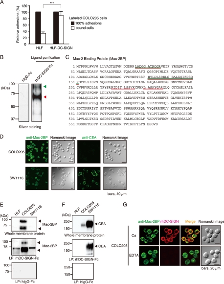
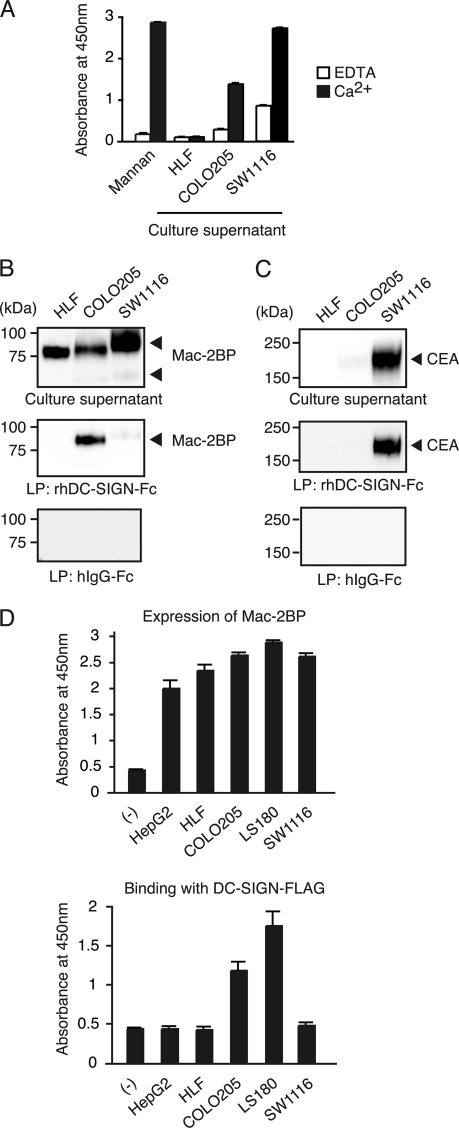
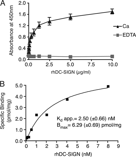
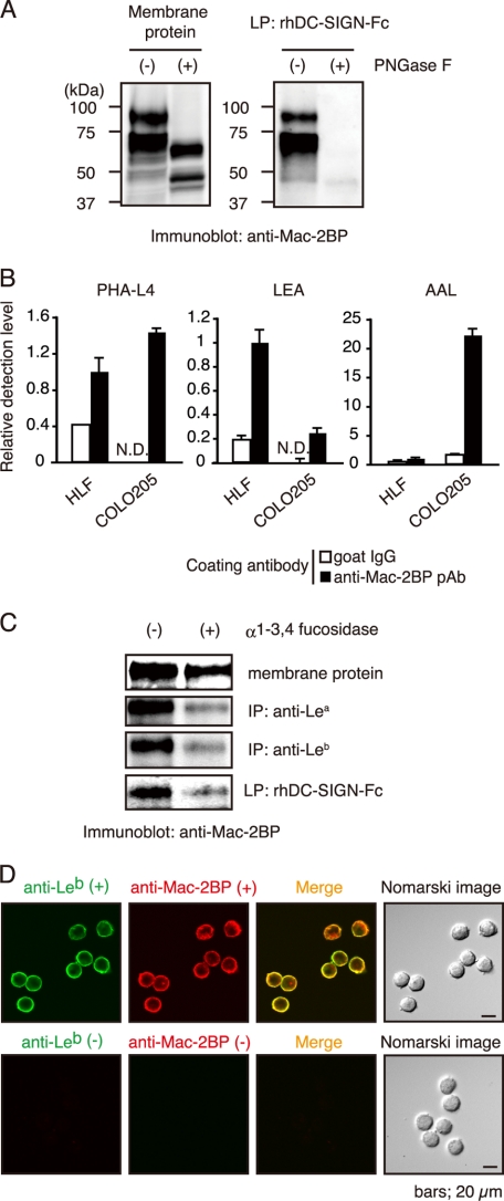
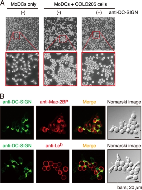
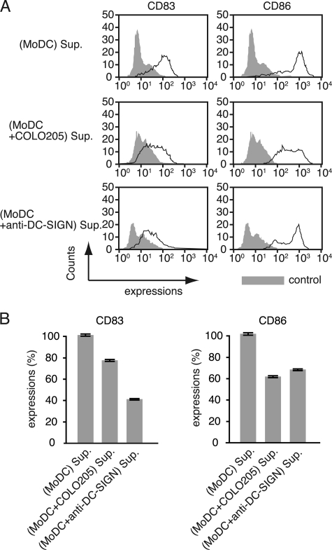
Dendritic cell (DC)-specific intercellular adhesion molecule-3-grabbing non-integrin (DC-SIGN) is a type II transmembrane C-type lectin expressed on DCs such as myeloid DCs and monocyte-derived DCs (MoDCs). Recently, we have reported that DC-SIGN interacts with carcinoembryonic antigen (CEA) expressed on colorectal carcinoma cells. CEA is one of the most widely used tumor markers for gastrointestinal cancers such as colorectal cancer. On the other hand, other groups have reported that the level of Mac-2-binding protein (Mac-2BP) increases in patients with pancreatic, breast, and lung cancers, virus infections such as human immunodeficiency virus and hepatitis C virus, and autoimmune diseases. Here, we first identified Mac-2BP expressed on several colorectal carcinoma cell lines as a novel DC-SIGN ligand through affinity chromatography and mass spectrometry. Interestingly, we found that DC-SIGN selectively recognizes Mac-2BP derived from some colorectal carcinomas but not from the other ones. Furthermore, we found that the α1-3,4-fucose moieties of Le glycans expressed on DC-SIGN-binding Mac-2BP were important for recognition. DC-SIGN-dependent cellular interactions between immature MoDCs and colorectal carcinoma cells significantly inhibited MoDC functional maturation, suggesting that Mac-2BP may provide a tolerogenic microenvironment for colorectal carcinoma cells through DC-SIGN-dependent recognition. Importantly, Mac-2BP was detected as a predominant DC-SIGN ligand expressed on some primary colorectal cancer tissues from certain parts of patients in comparison with CEA from other parts, suggesting that DC-SIGN-binding Mac-2BP bearing tumor-associated Le glycans may become a novel potential colorectal cancer biomarker for some patients instead of CEA.


Figures

FIGURE 1.
FIGURE 1.
Identification of Mac-2BP expressed on colorectal carcinomas as a novel ligand for DC-SIGN. A, DC-SIGN-mediated cellular adhesions with COLO205 cells. HLF or HLF-DC-SIGN cells bound with calcein-AM-labeled COLO205 cells were lysed and analyzed by fluorometry at 488 nm. The relative adhesions are shown. Error bars indicate S.D. (n = 3). B, purification of DC-SIGN ligands expressed on COLO205 cells. DC-SIGN ligands were purified with a DC-SIGN affinity column and then detected by silver staining. The arrowheads indicate the elution positions of the purified DC-SIGN ligands. The molecular mass markers are shown on the left. C, identification of DC-SIGN novel ligands by MS. The purified DC-SIGN ligand bands were analyzed by MS. The identified peptides are shown in green (upper band) or red (lower band), respectively. D, confocal microscopic images of the expressions of Mac-2BP and CEA on COLO205 and SW1116 cells. Cells were stained with anti-Mac-2BP pAb or anti-CEA mAb (green). Nomarski images are shown on the right. E and F, ligand precipitation (LP) analysis of the interaction of DC-SIGN with CEA or Mac-2BP. Solubilized membrane proteins of HLF, COLO205, and SW1116 cells were precipitated with rhDC-SIGN-Fc or hIgG-Fc, and then EDTA-eluted DC-SIGN ligands were detected by immunoblotting using anti-Mac-2BP pAb (E) or anti-CEA mAb (F). G, confocal microscopic images of the interactions between DC-SIGN and Mac-2BP on COLO205 cells. The cells were costained with anti-Mac-2BP pAb (green) and rhDC-SIGN-allophycocyanin (red) in the presence of CaCl2 or EDTA.
FIGURE 2.
FIGURE 2.
Culture supernatants derived from colorectal carcinoma cells contain secreted Mac-2BP that can be recognized by DC-SIGN. A, culture supernatant of colorectal carcinoma cells contain DC-SIGN-ligands. Total culture supernatant proteins from HLF, COLO205, and SW1116 cells, coated onto plates, were detected with rhDC-SIGN in the presence of 5 mm CaCl2 or EDTA. Mannan was used as a positive control for DC-SIGN binding. Error bars indicate S.D. (n = 3). B and C, DC-SIGN interacts selectively with COLO205-derived Mac-2BP or SW1116-derived CEA contained in culture supernatants. A total culture supernatant and purified DC-SIGN ligands from HLF, COLO205, or SW1116 cells were immunoblotted with anti-Mac-2BP pAb or anti-CEA mAb. D, colorectal carcinoma cells secrete Mac-2BP that can be recognized by DC-SIGN. Mac-2BPs from human hepatoma HepG2 and HLF and colorectal carcinoma COLO205, LS180, and SW1116 cells were captured with anti-Mac-2BP pAb and then detected with anti-Mac-2BP mAb (upper panel) or with rhDC-SIGN-FLAG (lower panel), respectively. Error bars indicate S.D. (n = 3).
FIGURE 3.
FIGURE 3.
Biochemical analysis of the interaction between DC-SIGN and Mac-2BP. A, DC-SIGN-dependent interaction with COLO205-derived Mac-2BP. Mac-2BP, captured with anti-Mac-2BP pAb onto plates, was detected with different concentrations of rhDC-SIGN-FLAG in the presence of 5 mm CaCl2 or EDTA. Error bars indicate S.D. (n = 3). B, quantitative kinetic analysis for association between DC-SIGN and Mac-2BP. The apparent (app.) dissociation constant (Kd app.) and Bmax were calculated based on the amounts of total and unbound rhDC-SIGN-FLAG using a nonlinear regression model.
FIGURE 4.
FIGURE 4.
Fucose residues of colorectal carcinoma-associated Lewis N-glycans expressed on COLO205-derived Mac-2BP are essential for DC-SIGN binding. A, N-glycans of COLO205-derived Mac-2BP are essential for DC-SIGN binding. COLO205 membrane proteins were incubated with or without PNGase F, and then total membrane proteins (left) and subsequently purified DC-SIGN-ligands (right) were immunoblotted with anti-Mac-2BP pAb. B, determination for the Mac-2BP glycan profile by ELISA. COLO205-derived Mac-2BP glycoproteins captured with pre-coated anti-Mac-2BP pAb were detected with biotinylated plant lectin PHA-L4 (left), Lycopersicon esculentum agglutinin (LEA, middle), or AAL (right). Goat IgG was used as an isotype control for anti-Mac-2BP capture pAb. Detection levels for HLF-derived Mac-2BP were arbitrarily set at 1. Error bars indicate S.D. (n = 3). C, DC-SIGN recognizes the fucose residues of Le glycans expressed on COLO205-derived Mac-2BP. COLO205 membrane proteins were incubated with or without α1-3,4-fucosidase and then precipitated with rhDC-SIGN-Fc, anti-Lea or -Leb mAb, followed by immunoblotting with anti-Mac-2BP pAb. IP, immunoprecipitation. D, confocal microscopic image of Leb expression on COLO205-derived Mac-2BP. The cells were costained with anti-Leb mAb (green) and anti-Mac-2BP pAb (red). Staining without primary Abs was performed as a negative control. LP, ligand precipitation.
FIGURE 5.
FIGURE 5.
DC-SIGN mediates interaction between MoDCs and COLO205 cells. A, DC-SIGN-dependent cellular adhesions between MoDCs and COLO205 cells. MoDCs only (left) or MoDCs plus COLO205 cells in the presence (right) or absence (middle) of anti-human DC-SIGN pAb were cultured and visualized by phase-contrast microscopy. B, confocal microscopic images of MoDC-COLO205 cellular interactions mediated by DC-SIGN and Mac-2BP. MoDCs were cocultured with COLO205 cells. After fixation, cells were stained with anti-human DC-SIGN mAb (green) and anti-Mac-BP pAb or anti-Leb mAb (red).
FIGURE 6.
FIGURE 6.
A COLO205-MoDC coculture-derived supernatant suppresses the functional maturation of MoDCs. A, flow cytometry analysis of CD83 and CD86 expressions. Immature MoDCs were incubated with a MoDC-cultured or COLO205-MoDC-cocultured supernatant (Sup.) for 3 days in the presence of IL-4, GM-CSF, and LPS (1 ng/ml), and the cultures were replenished with fresh supernatant on the second day. Supernatant of MoDCs, which cultured in the presence of anti-DC-SIGN pAb, was used as a control. The effect on MoDC functional maturation was determined as MoDC surface expression of CD83 and CD86 determined by flow cytometry. B, the relative expression levels of CD83 and CD86. The inhibition of MoDC functional maturation was measured as the percentage of mean fluorescent intensity (± S.E.) of incubation with MoDC-cultured supernatant. All experiments were performed in triplicate and were repeated a minimum of three times.
FIGURE 7.
FIGURE 7.
Mac-2BP expressed on colorectal cancer cells is a major target for DC-SIGN in situ. A, DC-SIGN recognizes colorectal carcinoma-derived Mac-2BP in situ. Primary colorectal cancer and normal colon tissues of donor 1 (SuperBioChips Laboratories) and donor 2 (Shiga University of Medical Science) were stained with rhDC-SIGN (green) and anti-Mac-2BP pAb (red). B, expressions of CEA and Mac-2BP in colorectal tumor tissues (SuperBioChips Laboratories). Triple staining of primary colorectal cancer and normal colon tissues with anti-CEA mAb (green), anti-Mac-2BP pAb (red), and rhDC-SIGN-allophycocyanin (blue). All samples were examined by confocal microscopy.

References

    1. Banchereau J., Briere F., Caux C., Davoust J., Lebecque S., Liu Y. J., Pulendran B., Palucka K. (2000) Annu. Rev. Immunol. 18, 767–811 - PubMed
    1. Lanzavecchia A., Sallusto F. (2001) Cell 106, 263–266 - PubMed
    1. Geijtenbeek T. B., van Vliet S. J., Engering A., ‘t Hart B. A., van Kooyk Y. (2004) Annu. Rev. Immunol. 22, 33–54 - PubMed
    1. van Vliet S. J., den Dunnen J., Gringhuis S. I., Geijtenbeek T. B., van Kooyk Y. (2007) Curr. Opin. Immunol. 19, 435–440 - PubMed
    1. van Kooyk Y., Rabinovich G. A. (2008) Nat. Immunol. 9, 593–601 - PubMed

Publication types

MeSH terms