. 2011 Jun 16;117(24):6608-11.
doi: 10.1182/blood-2010-12-322834. Epub 2011 Apr 22.

Defective inhibition of B-cell proliferation by Wiskott-Aldrich syndrome protein-deficient regulatory T cells

Affiliations

Defective inhibition of B-cell proliferation by Wiskott-Aldrich syndrome protein-deficient regulatory T cells

Marsilio Adriani et al. Blood. .

Abstract

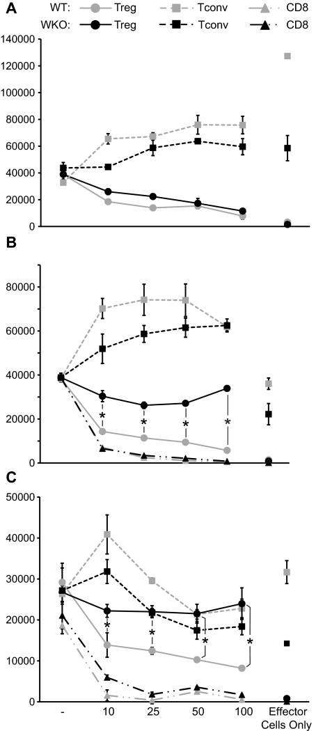
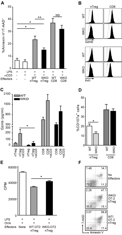
Wiskott-Aldrich syndrome (WAS) is an inherited immunodeficiency characterized by high incidence of autoantibody-mediated autoimmune complications. Such a feature has been associated with defective suppressor activity of WAS protein-deficient, naturally occurring CD4(+)CD25(+)Foxp3(+) regulatory T cells on responder T cells. However, it remains to be established whether the altered B-cell tolerance reported in WAS patients and Was knockout (WKO) mice is secondary to abnormalities in the direct suppression of B-cell function by nTreg cells or to impaired regulation of T-helper function. Because activated nTreg cells are known to induce granzyme B-mediated B-cell killing, we decided to evaluate the regulatory capabilities of WKO nTregs on B lymphocytes. We found that preactivated WKO nTreg cells failed to effectively suppress B-cell proliferation and that such a defect was associated with reduced killing of B cells and significantly decreased degranulation of granzyme B. Altogether, these results provide additional mechanistic insights into the loss of immune tolerance in WAS.


Figures

Figure 1
Figure 1
Preactivated WKO nTreg cells can suppress T-cell but not B-cell proliferation. nTreg, Tconv, and CD8+ T cells were isolated from WT and WKO mice and preactivated with anti-CD3 and IL-2. (A) Proliferation of freshly isolated Tconv (5 × 10⁴) from WT mice cocultured with the indicated number of WT and WKO preactivated nTreg or Tconv cells in the presence of anti-CD3 (2 μg/mL) and irradiated, T-depleted APCs. (B) B cells (5 × 10⁴) from WT mice were stimulated with LPS (3 μg/mL) and cocultured with the indicated number of preactivated nTreg, Tconv, or CD8+ cells from both WT or WKO mice in the presence of soluble anti-CD3 (2 μg/mL) and irradiated T-depleted APCs. (C) B cells isolated from WKO mice were cultured as in panels A and B. Proliferation was then assessed as incorporation of [3H]-thymidine after the cells were pulsed for the last 16 hours during a total of 72 hours of cultures. Note that the proliferation of effector T cells (nTreg, Tconv, and CD8 T cells) cultured alone in the presence of soluble anti-CD3 and LPS stimulation is also shown. Statistical significance of differences between samples was calculated using the 2-tailed Mann-Whitney test with 95% confidence intervals. Results are mean of triplicate cultures and are representative of at least 3 experiments. P < .05.
Figure 2
Figure 2
Defective B-cell death induction and granzyme B release by WKO nTreg cells. (A) Percentage of apoptotic B220+ cells after coculture of activated nTreg and CD8+ cells from WKO or WT mice with LPS-induced B-cell blasts at a ratio of 5:1 in the presence or absence of anti-CD3 antibody and LPS (0.5 μg/mL). Bar graph represents the results of 11 independent experiments expressed as mean ± SD. *P < .0001. **P < .05. NS indicates not significant. (B) Granzyme B (Gzmb) and perforin (Prf1) expression in WKO and WT preactivated nTreg and CD8+ cells. Open histograms represent isotype controls; and solid histograms, Gzmb- and Prf1-specific staining. Representative data from at least 3 independent experiments are shown. (C) ELISA determination of granzyme B (Gzmb) release in culture media by preactivated WT and WKO nTreg and CD8+ cells after restimulation with 2 μg/mL anti-CD3 antibody. Results are mean ± SD of 5 independent experiments. *P = .0260. Statistical significances of differences among samples were calculated using the 2-tailed Mann-Whitney test. (D) Percentage of CD107a+ after culture of WKO and WT-activated nTreg and CD8+ cells (5 × 10⁴) with 2 μg/mL soluble anti-CD3 and irradiated T cell–depleted spleen cells (5 × 10⁴) for 8 hours in the presence of FITC-anti–mouse CD107a or FITC-conjugated isotype control and GolgiStop (BD Biosciences PharMingen). After restimulation, the cells were stained with anti-CD107a, anti-CD4, and anti-CD8 and analyzed by flow cytometry. Results are mean ± SD of 3 independent experiments. *P = .0317. Statistical significances of differences among samples were calculated using the 2-tailed Mann-Whitney test. (E-F) Defective suppression of B-cell proliferation and LPS-induced B-cell blast killing by WKO-OT2 nTreg cells. (E) B cells (5 × 10⁴) from WT mice were stimulated with LPS (3 μg/mL) and cocultured with equal number of preactivated nTreg cells from both WT or WKO mice in the presence of OVA323-339 peptide (10μM), and irradiated T-depleted APCs. Proliferation was then assessed as in Figure 1. (F) B-cell blasts were pulsed with OVA323-339 peptide (10μM) for 2 hours and cultured alone or with WKO or WT OT-2 nTreg cells at an effector-to-target ratio of 1:1 for 8 hours in the presence of LPS (0.5 μg/mL). Statistical significance of differences between samples was calculated using the Mann-Whitney test with 95% confidence intervals. Results are mean of triplicate cultures and are representative of at least 2 experiments.

References

    1. Bosticardo M, Marangoni F, Aiuti A, Villa A, Grazia Roncarolo M. Recent advances in understanding the pathophysiology of Wiskott-Aldrich syndrome. Blood. 2009;113(25):6288–6295. - PubMed
    1. Ochs HD, Thrasher AJ. The Wiskott-Aldrich syndrome. J Allergy Clin Immunol. 2006;117(4):725–738. quiz 739. - PubMed
    1. Zhang J, Shehabeldin A, da Cruz LA, et al. Antigen receptor-induced activation and cytoskeletal rearrangement are impaired in Wiskott-Aldrich syndrome protein-deficient lymphocytes. J Exp Med. 1999;190(9):1329–1342. - PMC - PubMed
    1. Westerberg L, Larsson M, Hardy SJ, Fernandez C, Thrasher AJ, Severinson E. Wiskott-Aldrich syndrome protein deficiency leads to reduced B-cell adhesion, migration, and homing, and a delayed humoral immune response. Blood. 2005;105(3):1144–1152. - PubMed
    1. Humblet-Baron S, Sather B, Anover S, et al. Wiskott-Aldrich syndrome protein is required for regulatory T cell homeostasis. J Clin Invest. 2007;117(2):407–418. - PMC - PubMed

MeSH terms