Review
. 2011;48(1):1-35.
doi: 10.3233/BIR-2011-0579.

Biomechanics of leukocyte rolling

Affiliations
Review

Biomechanics of leukocyte rolling

Prithu Sundd et al. Biorheology. 2011.

Abstract

Leukocyte rolling on endothelial cells and other P-selectin substrates is mediated by P-selectin binding to P-selectin glycoprotein ligand-1 expressed on the tips of leukocyte microvilli. Leukocyte rolling is a result of rapid, yet balanced formation and dissociation of selectin-ligand bonds in the presence of hydrodynamic shear forces. The hydrodynamic forces acting on the bonds may either increase (catch bonds) or decrease (slip bonds) their lifetimes. The force-dependent 'catch-slip' bond kinetics are explained using the 'two pathway model' for bond dissociation. Both the 'sliding-rebinding' and the 'allosteric' mechanisms attribute 'catch-slip' bond behavior to the force-induced conformational changes in the lectin-EGF domain hinge of selectins. Below a threshold shear stress, selectins cannot mediate rolling. This 'shear-threshold' phenomenon is a consequence of shear-enhanced tethering and catch bond-enhanced rolling. Quantitative dynamic footprinting microscopy has revealed that leukocytes rolling at venular shear stresses (>0.6 Pa) undergo cellular deformation (large footprint) and form long tethers. The hydrodynamic shear force and torque acting on the rolling cell are thought to be synergistically balanced by the forces acting on tethers and stressed microvilli, however, their relative contribution remains to be determined. Thus, improvement beyond the current understanding requires in silico models that can predict both cellular and microvillus deformation and experiments that allow measurement of forces acting on individual microvilli and tethers.


Figures

Fig. 1
Fig. 1
In vitro rolling velocity of human neutrophils plotted as a function of P-selectin site density and wall shear stress. Data reproduced from Ref. [105]. Closed triangle (400 sites/μm2), closed square (200 sites/μm2), and closed circle (50 sites/ μm2).
Fig. 2
Fig. 2
Forces acting on a rolling neutrophil. Fs and Ts are the shear force (along x-axis) and torque (along y-axis about the cell center), respectively, exerted by the streaming fluid on the rolling cell. A single long tether ‘te1’ (representative of n tethers) and a stressed microvilli ‘mv1’ (representative of m stressed tetherless microvilli) are shown. The distance of the tether and microvillus bonds anchorage points from the center of the cell is denoted as Li (i = 1 to n) and lj (j = 1 to m), respectively. The tethers and the stressed microvilli are shown to form an angle φi (i = 1 to n) and θj (j = 1 to m) with the P-selectin substrate, respectively. The hydrodynamic force acting on the tethers and stressed tetherless microvilli is denoted as Fte,i (i = 1 to n) and Fmv,j (j = 1 to m), respectively. The cell radius is denoted by rc. The normal reaction force exerted by the substrate on the cell is denoted as FN. P-selectin on the substrate and PSGL-1 on the microvilli shown in blue and green color, respectively. The Cartesian coordinate system is shown in grey. vx(z) is the fluid velocity along x-axis. Not drawn to scale. Symbols of biophysical parameters are also explained in Table 2.
Fig. 3
Fig. 3
Stressed, compressed, and unstressed bonds in the footprint of a rolling neutrophil [3D-reconstruction adapted from Sundd, P. et al. Nat Methods 7, 821-824 (2010) with permission]. 3D reconstruction of the footprint of an EGFP-expressing neutrophil rolling on P-selectin coated substrate generated using qDF microscopy. A microvillus is defined as a conical protrusion longer than 25 nm in z-direction. Only three microvilli (mv1, mv2, and mv3) are labeled. Schematics of P-selectin-PSGL-1 molecular complex (not to scale) drawn on the tips of microvilli. As the cell rolls from left to right, the P-selectin-PSGL-1 bonds are formed in the front at the unstressed length (Lsep ~ 70 nm; mv3), compressed in the center (Lsep < 70 nm; mv2) and stressed beyond the unstressed length (Lsep > 70 nm; mv3) at the rear of the footprint where they finally break. P-selectin and PSGL-1 shown as blue and green, respectively. Wall shear stress 0.6 Pa. P-selectin 20 molecules/μm2.
Fig. 4
Fig. 4
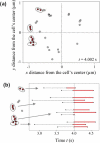
ETMA simulations reveal bond clusters in the footprint of a rolling neutrophil. (a) Distribution of the PSGL-1-P-selectin bond bases in the footprint of a rolling neutrophil (the cell translates in the positive x-direction). The load-bearing bonds are marked with red circles. Ovals indicate the load-bearing bond clusters and each cluster corresponds to a different microvillus. (b) Lifetime of 11 load-bearing bonds seen in (a). Each bond is represented by a black segment (the no-load time period) and a red segment (the load time period). All bonds but one become load-bearing at the same time (t = 4 s). Unpublished data (Pospieszalska and Ley).
Fig. 5
Fig. 5
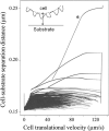
Scatter plot showing cell translational velocity as a function of cell body to substrate seperation distance (distance does not include microvilli-see inset). Instantaneous changes in the separation distance are seen as individual traces. The asterisk indicates the trace representing the first approach of the cell to the substrate. The almost vertical trace for the high translational velocities represents instantaneous changes in the separation distance when no load-bearing bonds were present. The trace discontinuity near velocity zero is due to a discrete process of modeling owing to the time step. Unpublished data (Pospieszalska and Ley).
Fig. 6
Fig. 6
Whole cell deformation results in a larger footprint at high shear stress [(b) and (c) adapted from Sundd, P. et al. Nat Methods 7, 821-824 (2010) with permission]. (a) A schematic showing the side and bottom views of the footprint of a rolling neutrophil following deformation by the shear forces to a nonspherical shape. Dashed circle shows the nondeformed neutrophil for comparison. Red closed circles are the tips of microvilli in the footprint. (b) Coordinates of the microvilli tips in the footprint of an EGFP-expressing neutrophil rolling on P-selectin substrate extracted from qDF micrographs. P-selectin 20 molecules/μm2. Wall shear stress 0.6 Pa. (c) Coordinates of the microvilli tips in the footprint of a non-deformable spherical rolling neutrophil revealed using ETMA simulations. P-selectin 150 molecules/μm2. Wall shear stress 0.05 Pa. Color of the data points in (b) and (c) represents the z-coordinates of microvilli tips based on color bars shown on the right. Black arrows denote the direction of rolling.
Fig. 7
Fig. 7
Microvillus deformation under tensile force. Schematic showing a neutrophil interacting with a P-selectin coated substrate through PSGL-1 expressed on the microvillus tip. To denote the individual microvillus/tether force acting on the cell, symbol Fmv is used when there is no tether and symbol Fte when a tether is present. (a) Unstressed microvillus for Fmv = 0. (b) Stressed microvillus without tether for 0 < Fmv < Fth. (c) Stressed microvillus with a long membrane tether formed following dissociation of plasma membrane from the cytoskeleton under force Fte = Fmv > Fth. The symbols for biophysical parameters are defined in Table 2. P-selectin and PSGL-1 shown as blue and green, respectively. Actin cytoskeleton shown in red. Tether anchorage point shown as a round structure at the end of tether. Not drawn to scale.
Fig. 8
Fig. 8
Models for the microvillus extension material used in published PSGL-1/P-selectin pulling experiments. (a) Elastic model represented by a spring. (b) Kelvin-Voigt viscoelastic model represented by a spring and a dashpot connected in parallel. (c) Viscoelastic model composed of a Kelvin-Voigt unit and an elastic unit connected in series. In a-c, σmv and κmv are the spring constants for the corresponding springs, ηeff_mv is the effective viscosity of the dashpot, Fmv is the pulling force, Lmv is the microvillus extension, mv = dLmv/dt is the microvillus extension rate, and mv = dFmv/dt is the loading rate. (d) Microvillus extension under a constant force, Fmv = 45 pN, described by the models a-c for σmv = 43 pN/μm, = ηeff_mv = 33 pN s/μm, and κmv = 800 pN/μm.
Fig. 9
Fig. 9
Long tethers facilitate neutrophil rolling at high shear stress [Adapted from Sundd, P. et al. Nat Methods 7, 821-824 (2010) with permission]. (a) qDF micrograph of a DiI-labeled neutrophil rolling on P-selectin substrate processed to saturation to reveal anchorage points of long tethers behind the footprint. (b) 3D reconstruction of the footprint shown in (a) reveals z position of tether anchorage points above the P-selectin substrate which is equal to the length of the stressed P-selectin-PSGL-1 bonds holding the tether anchorage point to the substrate. P-selectin 20 molecules/μm2. Wall shear stress 0.8 Pa. Flow is from left to right. Scale bar 10 μm. Arrows mark tether anchorage points.

References

    1. Addison W. Experimental and practical researches on the structure and function of blood corpuscles; on inflammation, and on the origin and nature of tubercules in the lungs. Trans. Provinc. Med. Surg. Ass. 1843;11:223–306.
    1. Afshar-Kharghan V, Diz-Kucukkaya R, Ludwig EH, et al. Human polymorphism of P-selectin glycoprotein ligand 1 attributable to variable numbers of tandem decameric repeats in the mucinlike region. Blood. 2001;97:3306–3307. - PubMed
    1. Alberts B, Bray D, Lewis J, et al. Molecular biology of the cell. Garland Publishing, Inc.; New York: 1994.
    1. Alon R, Chen S, Fuhlbrigge R, et al. The kinetics and shear threshold of transient and rolling interactions of L-selectin with its ligand on leukocytes. Proc. Natl. Acad. Sci. USA. 1998;95:11631–11636. - PMC - PubMed
    1. Alon R, Chen S, Puri KD, et al. The kinetics of L-selectin tethers and the mechanics of selectin-mediated rolling. J. Cell Biol. 1997;138:1169–1180. - PMC - PubMed

Publication types

Substances

LinkOut - more resources