

Change in mean transit time, apparent diffusion coefficient, and cerebral blood volume during pediatric diabetic ketoacidosis treatment
- PMID: 21516055
- PMCID: PMC3157541
- DOI: 10.1097/PCC.0b013e3182196c9c
Change in mean transit time, apparent diffusion coefficient, and cerebral blood volume during pediatric diabetic ketoacidosis treatment
Abstract
Objectives: Cerebral edema is a devastating complication of pediatric diabetic ketoacidosis. We examined measures describing potential causes of whole brain and regional brain edema (mean transit time, apparent diffusion coefficient, and relative cerebral blood volume) during treatment of diabetic ketoacidosis in children.
Design: Prospective observational study.
Setting: Regional children's hospital.
Interventions: None.
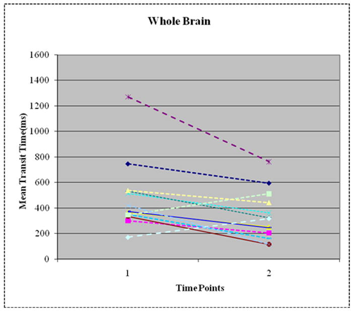
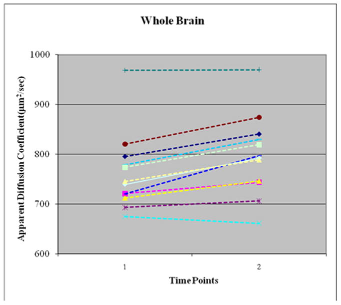
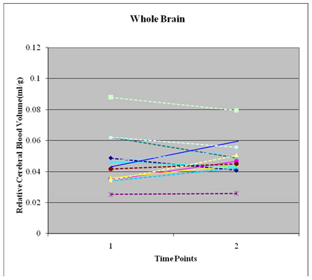
Measurements and main results: After Institutional Review Board approval, children admitted with diabetic ketoacidosis (pH <7.3, HCO3 <15 mEq/L, glucose >300 mg/dL, and ketosis) underwent two serial paired contrast-enhanced (gadolinium) and diffusion magnetic resonance imaging scans. Change in whole brain and regional (frontal lobe, occipital lobe, and basal ganglia) mean transit time, apparent diffusion coefficient, and relative cerebral blood volume between the two time periods (12-24 hrs) and (36-72 hrs) after start of insulin treatment (time 0) were determined. Thirteen children (median age, 10.3 ± 1.1 yrs; 7 female) with diabetic ketoacidosis were examined. Overall, whole brain and regional mean transit time decreased from time 1 (first magnetic resonance imaging after time 0) to time 2 (second magnetic resonance imaging after time 0) by 51% ± 59% (p = .01), without differences between the brain regions examined. Whole brain apparent diffusion coefficient increased by 4.7% ± 3.4% (p = .001), without differences between the brain regions examined. There was no change in relative cerebral blood volume for the whole brain and for the three brain regions examined.
Conclusions: In this study, whole brain mean transit time decreased and apparent diffusion coefficient increased, suggesting a vasogenic process between the two study periods during diabetic ketoacidosis treatment.
Figures
Comment in
-
Cerebral edema in diabetic ketoacidosis: time to go with the (cerebral blood) flow?Pediatr Crit Care Med. 2011 Nov;12(6):687-9. doi: 10.1097/PCC.0b013e3182231248. Pediatr Crit Care Med. 2011. PMID: 22067822 No abstract available.
References
-
- Rewers A, Chase HP, Mackenzie T, et al. Predictors of acute complications in children with type 1 diabetes. JAMA. 2002;287:2511–2518. - PubMed
-
- Rewers A, Klingensmith G, Davis C, et al. Presence of diabetic ketoacidosis at diagnosis of diabetes mellitus in youth: the Search for Diabetes in Youth Study. Pediatrics. 2008;121:e1258–66. - PubMed
-
- Hanas R, Lindblad B, Lindgren F. Diabetic ketoacidosis and cerebral edema in Sweden, a 2-year population study (Abstract) Diabetes. 2004;53 (Suppl 2):A421. - PubMed
-
- Roche EF, Menon A, Gill D, et al. Clinical presentation of type 1 diabetes. Pediatr Diabetes. 2005;6:75–78. - PubMed
-
- Edge JA. Cerebral oedema during treatment of diabetic ketoacidosis: are we any nearer finding a cause? Diabetes Metab Res Rev. 2000;16:316–324. - PubMed
Publication types
MeSH terms
Grants and funding
LinkOut - more resources
Full Text Sources
