. 2011 Jun;12(6):568-75.
doi: 10.1038/ni.2031. Epub 2011 Apr 24.

The encephalitogenicity of T(H)17 cells is dependent on IL-1- and IL-23-induced production of the cytokine GM-CSF

Affiliations

The encephalitogenicity of T(H)17 cells is dependent on IL-1- and IL-23-induced production of the cytokine GM-CSF

Mohamed El-Behi et al. Nat Immunol. 2011 Jun.

Abstract

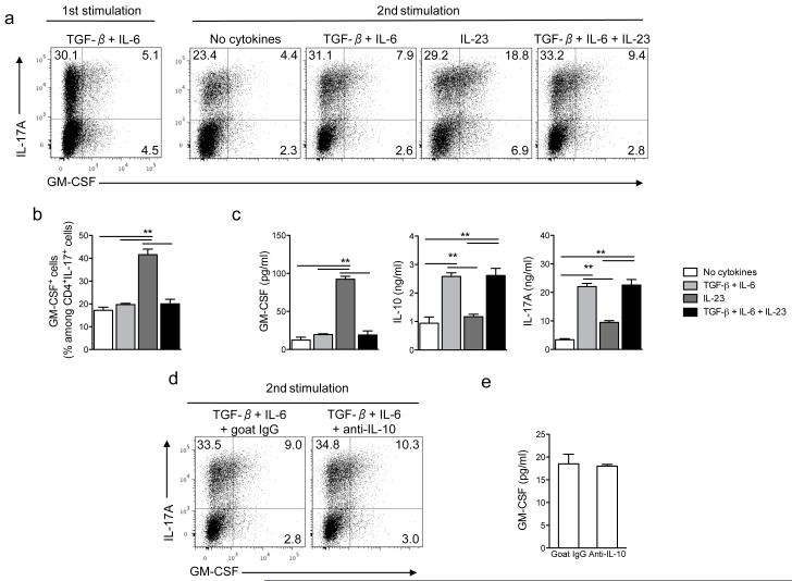
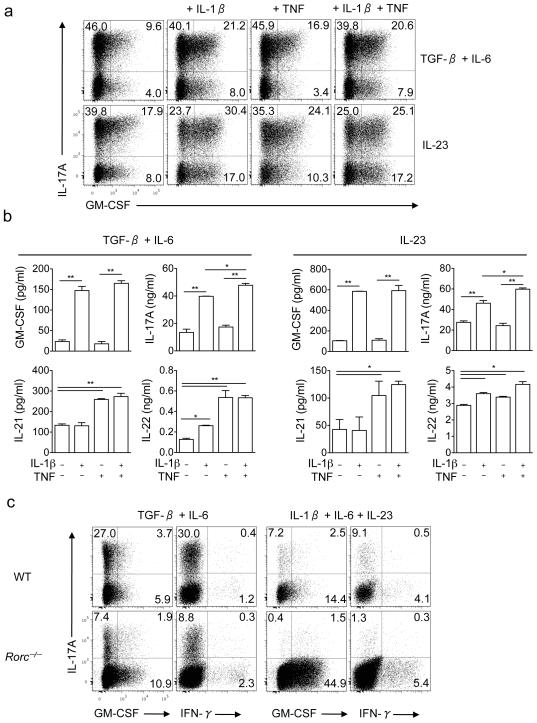
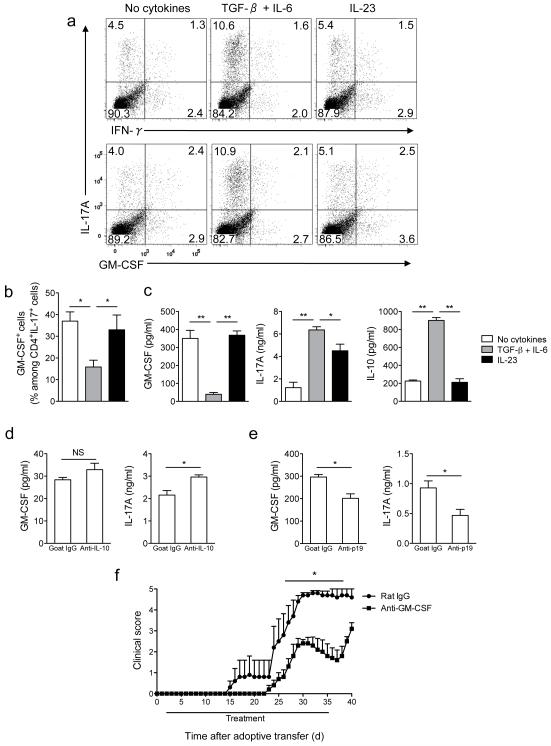
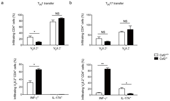
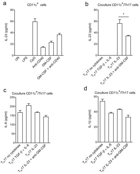
Interleukin 17 (IL-17)-producing helper T cells (T(H)17 cells) require exposure to IL-23 to become encephalitogenic, but the mechanism by which IL-23 promotes their pathogenicity is not known. Here we found that IL-23 induced production of the cytokine granulocyte-macrophage colony-stimulating factor (GM-CSF) in T(H)17 cells and that GM-CSF had an essential role in their encephalitogenicity. Our findings identify a chief mechanism that underlies the important role of IL-23 in autoimmune diseases. IL-23 induced a positive feedback loop whereby GM-CSF secreted by T(H)17 cells stimulated the production of IL-23 by antigen-presenting cells. Such cross-regulation of IL-23 and GM-CSF explains the similar pattern of resistance to autoimmunity when either of the two cytokines is absent and identifies T(H)17 cells as a crucial source of GM-CSF in autoimmune inflammation.


Figures

Figure 1
Figure 1. IL-23 upregulates GM-CSF expression in TH17 cells
(a) Naive CD4+CD25CD62LhiCD44lo T cells from spleens of C57BL/6 mice were sorted by flow cytometry and activated with anti-CD3 and anti-CD28 in the presence of TGF-β plus IL-6, anti-IFN-γ and anti-IL-4 for 72 h (first stimulation). Cells were rested 2 days in the presence of IL-2 and then reactivated with anti-CD3 and anti-CD28 (second stimulation) during 72 h in the presence of TGF-β plus IL-6, TGF-β, IL-6 and IL-23, IL-23 or without added cytokines. Cells were then stimulated with PMA and ionomycin in the presence of GolgiPlug for the final 4 h, stained and analyzed by flow cytometry. CD4+ cells are shown. (b) Percentage of GM-CSF+ cells among CD4+IL-17A+ cells after the second stimulation. (c) GM-CSF, IL-10 and IL-17A concentrations in cell culture supernatants after the second stimulation measured by ELISA. (d) TH17 cells restimulated in the presence of TGF-β and IL-6 for 72 h were treated with anti-IL-10 or isotype control (goat IgG) and analyzed by flow cytometry. (e) GM-CSF concentrations in culture supernatants of cells stimulated as in (d) measured by ELISA. **p< 0.001. Data are representative of three independent experiments. (error bars, s.e.m).
Figure 2
Figure 2. IL-1β upregulates GM-CSF expression in TH17 cells
(a) Naive CD4+CD25CD62LhiCD44lo T cells from spleens of C57BL/6 mice were FACS sorted and differentiated into TH17 cells during the first stimulation. Cells were then reactivated with anti-CD3 and anti-CD28 for 72 h in the presence of either IL-23 or TGF-β plus IL-6, in the presence of IL-1β (10 ng/ml) and/or TNF (10 ng/ml). CD4+ cells are shown. (b) GM-CSF, IL-17A, IL-21 and IL-22 concentrations in cell culture supernatants after the second stimulation measured by ELISA. (c) Naive CD4+ T cells from spleens of C57BL/6 and RORγt-deficient mice were activated during 72 h with anti-CD3 and anti-CD28 in the presence of either TGF-β and IL-6 or IL-1β, IL-6 and IL-23. Cells were then stimulated with PMA and ionomycin in the presence of GolgiPlug, stained and analyzed by flow cytometry. *p<0.01, **p < 0.001. Data are representative of two independent experiments. (error bars, s.e.m).
Figure 3
Figure 3. Expression of TH17 cells markers is regulated by IL-1β and TNF
Real-time PCR analysis of Ahr, CCR6, CCL20, IL-23R and RORα in TH17 cells reactivated with IL-23, TGF-β plus IL-6 or both in the presence of IL-1β (10 ng/ml) and/or TNF (10 ng/ml). *p < 0.01; **p < 0.001. Data are representative of two independent experiments. (error bars, s.e.m).
Figure 4
Figure 4. Neutralization of GM-CSF attenuates adoptive EAE
(a) Splenocytes from MOG35-55-immuzed C57BL/6 mice were activated with MOG35-55 (20 μg/ml) in the presence of TGF-β plus IL-6, IL-23 or without added cytokines. Cells were then stimulated with PMA and ionomycin in the presence of GolgiPlug, stained and analyzed by flow cytometry. CD4+ cells are shown. (b) Percentage of GM-CSF+ cells among CD4+IL-17A+ cells after the second stimulation. (c) GM-CSF, IL-10, and IL-17A concentrations in cell culture supernatants measured by ELISA. (d) GM-CSF and IL-17A concentrations in supernatants of cell cultures stimulated as in (a) in the presence of TGF-β plus IL-6 and treated with anti-IL-10 or goat IgG. (e) GM-CSF and IL-17A concentrations in supernatants of cell cultures stimulated as in (a) without added cytokines and treated with anti-IL-23p19 or goat IgG (f) Clinical scores of irradiated wild-type recipient mice that received 5×10⁶ CD4+ cells enriched from EAE splenocytes activated in the presence of IL-23. Mice were treated with either anti-GM-CSF or rat IgG from day 2 to day 35 post cell transfer. *p< 0.01; **p < 0.001. Data are representative of two (d, e and f) or four (a, b, and c) independent experiments (error bars, s.e.m).
Figure 5
Figure 5. GM-CSF production by TH1 and TH17 cells is required for their encephalitogenicity
Splenocytes from wild-type- or Csf2−/−-MBPAc1-11 transgenic mice were activated with MBPAc1-11 peptide in the presence of IL-12 (TH1 conditions) or TGF-β plus IL-6, anti-IFN-γ and anti-IL-4 during 72 h (TH17 conditions). Cells were rested 2 days in the presence of IL-2 and then reactivated with MBPAc1-11 peptide in the presence of IL-12 (TH1 conditions) or IL-23 (TH17 conditions). After 72 h, cells were stimulated with PMA and ionomycin in the presence of GolgiPlug. (a) Flow cytometric analysis of IL-17A and IFN-γ expression in wild-type and Csf2−/− TH1 and TH17 cells after the second stimulation. CD4+ cells are shown. (b) IL-17A, IFN-γ and GM-CSF concentrations were measured by ELISA in the supernatants after the second stimulation. (c) RORγt and T-bet expression on gated CD4+IFN-γ+ (TH1) or CD4+IL-17A+ cells (TH17) analyzed by flow cytometry after the second stimulation. Filled histograms represent isotype controls. (d) Real time PCR analysis of Ahr, CCL20, CCR6, IL-23R and RORα expression in CD4+ T cells enriched by magnetic beads after the second stimulation from wild-type or Csf2−/− MBPAc1-11-splenocytes cultured in TH17 conditions. (e) Clinical scores of recipient mice that received 5×10⁶ of either MBPAc1-11-specific wild-type or Csf2−/− TH1 or TH17 cells enriched by magnetic beads after the second stimulation. Pertussis toxin was injected i.p. on days 0 and 2 post transfer. *p< 0.01; **p < 0.001. Data are representative of two independent experiments. (error bars, s.e.m).
Figure 6
Figure 6. Csf2−/− TH1 and TH17 cells infiltrate the CNS but do not induce EAE
EAE was induced as described in Figure 5. Spinal cords of TH1 (a) and TH17 cells (b) recipient mice were harvested at day 25 p.i. and mononuclear cells were isolated and stimulated with PMA, ionomycin, and GolgiPlug. Cells were then analyzed by flow cytometry for the expression of CD4, Vβ8.2 (upper panels), IL-17A and IFN-γ (lower panels). *p< 0.01; **p < 0.001. Data are representative of two independent experiments. (error bars, s.e.m).
Figure 7
Figure 7. GM-CSF secreted by TH17 cells augments production of IL-23 by CD11c+ cells
(a) CD11c+ cells isolated from splenocytes of C57BL/6 mice were stimulated with LPS, CpG, anti-CD40, rGM-CSF or left untreated (UN) for 24 h. (b), (c) and (d) Differentiated OT-II TH17 cells were cultured overnight in the presence of indicated cytokines, washed extensively, and then cocultured with CD11c+ cells and OVA323-339. Cytokine concentrations in supernatants were measured by ELISA. *p < 0.01. Data are representative of two independent experiments (error bars, s.e.m).

Comment in

References

    1. Goverman J. Autoimmune T cell responses in the central nervous system. Nat Rev Immunol. 2009;9:393–407. - PMC - PubMed
    1. Bettelli E, et al. Reciprocal developmental pathways for the generation of pathogenic effector TH17 and regulatory T cells. Nature. 2006;441:235–238. - PubMed
    1. Elyaman W, et al. IL-9 induces differentiation of TH17 cells and enhances function of FoxP3+ natural regulatory T cells. Proc Natl Acad Sci U S A. 2009;106:12885–12890. - PMC - PubMed
    1. Korn T, et al. IL-21 initiates an alternative pathway to induce proinflammatory T(H)17 cells. Nature. 2007;448:484–487. - PMC - PubMed
    1. Mangan PR, et al. Transforming growth factor-beta induces development of the T(H)17 lineage. Nature. 2006;441:231–234. - PubMed

Publication types

MeSH terms