

Melanin-concentrating hormone: a new sleep factor?
- PMID: 21516258
- PMCID: PMC3080035
- DOI: 10.3389/fneur.2011.00014
Melanin-concentrating hormone: a new sleep factor?
Abstract
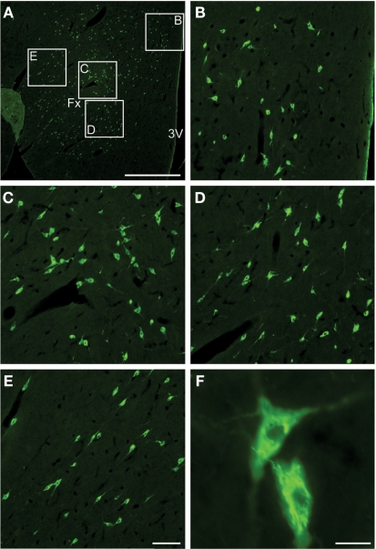
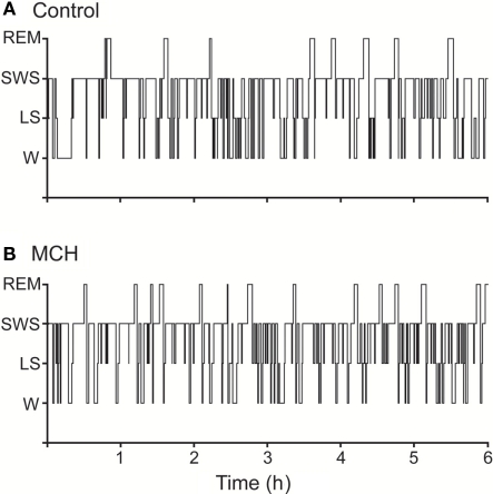
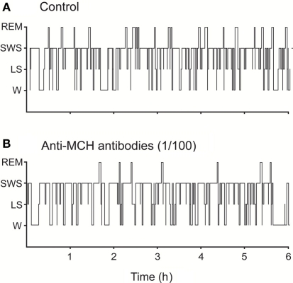
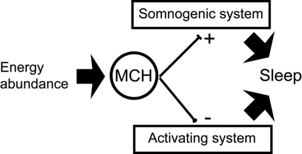
Neurons containing the neuropeptide melanin-concentrating hormone (MCH) are mainly located in the lateral hypothalamus and the incerto-hypothalamic area, and have widespread projections throughout the brain. While the biological functions of this neuropeptide are exerted in humans through two metabotropic receptors, the MCHR1 and MCHR2, only the MCHR1 is present in rodents. Recently, it has been shown that the MCHergic system is involved in the control of sleep. We can summarize the experimental findings as follows: (1) The areas related to the control of sleep and wakefulness have a high density of MCHergic fibers and receptors. (2) MCHergic neurons are active during sleep, especially during rapid eye movement (REM) sleep. (3) MCH knockout mice have less REM sleep, notably under conditions of negative energy balance. Animals with genetically inactivated MCHR1 also exhibit altered vigilance state architecture and sleep homeostasis. (4) Systemically administered MCHR1 antagonists reduce sleep. (5) Intraventricular microinjection of MCH increases both slow wave sleep (SWS) and REM sleep; however, the increment in REM sleep is more pronounced. (6) Microinjection of MCH into the dorsal raphe nucleus increases REM sleep time. REM seep is inhibited by immunoneutralization of MCH within this nucleus. (7) Microinjection of MCH in the nucleus pontis oralis of the cat enhances REM sleep time and reduces REM sleep latency. All these data strongly suggest that MCH has a potent role in the promotion of sleep. Although both SWS and REM sleep are facilitated by MCH, REM sleep seems to be more sensitive to MCH modulation.
Keywords: REM sleep; depression; hypothalamus; orexin; peptides; raphe nuclei.
Figures
References
-
- Adamantidis A., Salvert D., Goutagny R., Lakaye B., Gervasoni D., Grisar T., Luppi P. H., Fort P. (2008). Sleep architecture of the melanin-concentrating hormone receptor 1-knockout mice. Eur. J. Neurosci. 27, 1793–1800 - PubMed
-
- Adrien J. (2002). Neurobiological bases for the relation between sleep and depression. Sleep Med. Rev. 6, 341–351 - PubMed
-
- Ahnaou A., Drinkenburg W. H., Bouwknecht J. A., Alcazar J., Steckler T., Dautzenberg F. M. (2008). Blocking melanin-concentrating hormone MCH(1) receptor affects rat sleep-wake architecture. Eur. J. Pharmacol. 579, 177–188 - PubMed
-
- Astrand A., Bohlooly Y. M., Larsdotter S., Mahlapuu M., Andersen H., Tornell J., Ohlsson C., Snaith M., Morgan D. G. (2004). Mice lacking melanin-concentrating hormone receptor 1 demonstrate increased heart rate associated with altered autonomic activity. Am. J. Physiol. Regul. Integr. Comp. Physiol. 287, R749–R758 - PubMed
LinkOut - more resources
Full Text Sources
Miscellaneous