. 2011 Sep;106(5):829-47.
doi: 10.1007/s00395-011-0181-0. Epub 2011 Apr 24.

In vitro epithelial-to-mesenchymal transformation in human adult epicardial cells is regulated by TGFβ-signaling and WT1

Affiliations

In vitro epithelial-to-mesenchymal transformation in human adult epicardial cells is regulated by TGFβ-signaling and WT1

Noortje A M Bax et al. Basic Res Cardiol. 2011 Sep.

Abstract

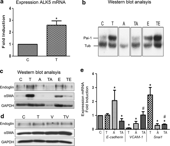
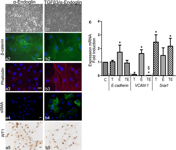
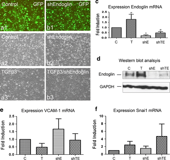
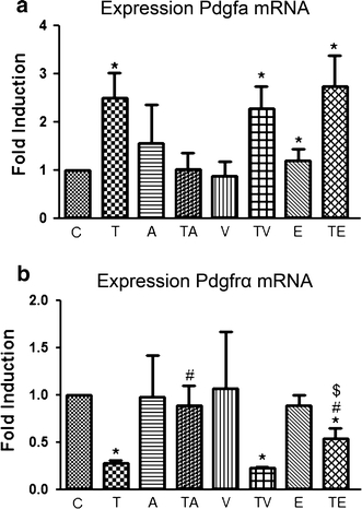
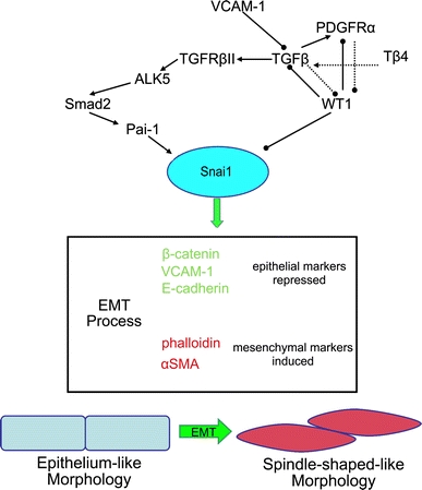
Adult epicardial cells are required for endogenous cardiac repair. After myocardial injury, they are reactivated, undergo epithelial-to-mesenchymal transformation (EMT) and migrate into the injured myocardium where they generate various cell types, including coronary smooth muscle cells and cardiac interstitial fibroblasts, which contribute to cardiac repair. To understand what drives epicardial EMT, we used an in vitro model for human adult epicardial cells. These cells have an epithelium-like morphology and markedly express the cell surface marker vascular cell adhesion marker (VCAM-1). In culture, epicardial cells spontaneously undergo EMT after which the spindle-shaped cells now express endoglin. Both epicardial cells before and after EMT express the epicardial marker, Wilms tumor 1 (WT1). Adding transforming growth factor beta (TGFβ) induces loss of epithelial character and initiates the onset of mesenchymal differentiation in human adult epicardial cells. In this study, we show that TGFβ-induced EMT is dependent on type-1 TGFβ receptor activity and can be inhibited by soluble VCAM-1. We also show that epicardial-specific knockdown of Wilms tumor-1 (WT1) induces the process of EMT in human adult epicardial cells, through transcriptional regulation of platelet-derived growth factor receptor alpha (Pdgfrα), Snai1 and VCAM-1. These data provide new insights into the process of EMT in human adult epicardial cells, which might provide opportunities to develop new strategies for endogenous cell-based cardiac repair.


Figures

Fig. 1
Fig. 1
Surface marker profile of human adult epicardial cells. EPDC cultures were stained using WT1 to certify the purity of the culture (a, b). Flow cytometric analysis of cultured adult human EPDCs before (cEPDCs) and after EMT (sEPDCs) was performed (c). Histograms of endoglin (CD105) (d) and VCAM-1 (CD106) (e) are shown with isotype control (dashed line) and the specific signal (solid line). Scale bar 20 μm
Fig. 2
Fig. 2
Western blot and qRT-PCR analysis. Gene expression of cEPDCs (control; C) and cEPDCs treated with 1 ng/ml TGFβ3 (T) showed that the expression of ALK5 was increased by TGFβ (a). Western blot analysis of protein samples isolated from cEPDCs stimulated in the presence or absence of 1 ng/ml TGFβ3 and α-Endoglin probed for Pai-1 (b). As a loading control, α/β-tubulin was used (b). Western blot analysis of αSMA and endoglin in epicardial cells stimulated by sVCAM-1 (100 ng/ml) and α-Endoglin (0.5 μg/ml) independent or simultaneously with TGFβ3 (1 ng/ml) (c, d). GAPDH was used as a loading control in this experiment (c, d). Gene expression of cEPDCs and cEPDCs treated with TGFβ3, iALK5 and both simultaneously. Treatment with TGFβ showed significant decrease in epithelial markers and increase in EMT marker Snai1, which is dependent on ALK5 kinase activity (e). *P < 0.05 versus control, # P < 0.05 versus TGFβ3 stimulation. For patterns and abbreviations see Box 1 in Appendix
Fig. 3
Fig. 3
TGFβ-stimulated EMT is dependent on ALK5 kinase activity. Transforming growth factor (TGFβ) induces epithelial-to-mesenchymal transformation (EMT) in human adult epicardial cells, and ALK5 is required for the effects of TGFβ. Epicardial cells were treated with 1 ng/ml TGFβ3, 10 μM SB431542 (iALK5) and ALK5 kinase inhibitor for 48 h before fixation. Untreated epicardial cells display an epithelial phenotype (a) accompanied with expression of β-catenin (a2) and phalloidin (a3) at the cell–cell borders. Cells incubated with TGFβ are elongated, have lost β-catenin expression and phalloidin was visualized across the cells in stress fibers (b13). Cells treated with TGFβ express αSMA consistent with a smooth muscle phenotype (b4). In the presence of iALK5, cells display an epithelial phenotype (c) consistent with the untreated cells (a). Cells treated simultaneously with TGFβ and iALK5 retain expression of β-catenin at the cell border and no phalloidin staining across the cells is present (d). All untreated and treated cells express WT1 (a5d5). ×100 in a1d1. Scale bar 20 μm
Fig. 4
Fig. 4
sVCAM-1 inhibits cell shape changes in human adult epicardial cells treated with TGFβ3. Unstimulated cells (a) and stimulated with sVCAM-1 (100 ng/ml) (b) and simultaneously with TGFβ3 (1 ng/ml) and sVCAM-1 (100 ng/ml) (c) are stained for β-catenin (a2c2) and with phalloidin to visualize filamentous actin (a3c3). Onset of differentiation into smooth muscle cells was visualized by staining for αSMA (a4c4) and the state of differentiation was visualized by WT1 (a5c5). The process of EMT was confirmed by qRT-PCR (d) analysis for epithelial and EMT markers. ×100 in a1c1. Scale bar 20 μm. *P < 0.05 versus control, # P < 0.05 versus TGFβ3 stimulation, $ P < 0.05 stimuli versus simultaneous TGFβ3/stimuli. For patterns and abbreviations see Box 1 in Appendix
Fig. 5
Fig. 5
Inhibition of endoglin expression cannot block the process of TGFβ-stimulated EMT. Blocking of endoglin increased the expression of epithelial markers (a, c). In simultaneous treatment of epicardial cells with TGFβ and α-Endoglin, the decrease in epithelial and increase in mesenchymal and EMT markers stimulated by TGFβ could not be prevented (ac). *P < 0.05 versus control, $ P < 0.05 stimuli versus simultaneous TGFβ3/stimuli. ×100 in a1b1. Scale bar 20 μm. For patterns and abbreviations see Box 1 in Appendix
Fig. 6
Fig. 6
Knockdown of endoglin expression cannot block the process of TGFβ-stimulated EMT. Transduction of epicardial cells with lentivirus expression shRNAs for control GFP virus (Control) and human endoglin (shEndoglin) was visualized by the expression of GFP (a1, b1) and did not affect the cell morphology (a2, b2). Addition of TGFβ caused morphological changes in both shEndoglin-transduced cEPDCs and control (a3, b3). qRT-PCR (c) and Western blot (d) analysis showed reduction of endogenous endoglin expression after transduction (c, d). Addition of TGFβ caused increase in endoglin expression in both transduced and control epicardial cells. Analysis of epithelial marker VCAM-1 (e) and EMT marker Snai1 (f) showed that knockdown of endoglin could not prevent the effects of TGFβ on these markers. ×100 in a1c2. *P < 0.05 versus control, # P < 0.05 TGFβ3 stimulation versus nonstimulated. C control, T TGFβ3; shE shEndoglin, shTE TGFβ3/shEndoglin
Fig. 7
Fig. 7
Role of WT1 in the EMT process of human adult epicardial cells. Quantification of WT1 isoform A mRNA expression in human adult epicardial cells decreased after treatment with TGFβ3 (a). Knockdown of WT1 by shRNA results in loss of epithelial character of epicardial cells (bd). Quantification of WT1 mRNA expression was significantly reduced in epicardial cells after knockdown of WT1 (P < 0.05) (e and Online Resource 3). The expression of epithelial markers E-cadherin (f), α4-integrin (g) and VCAM-1 (h) mRNA was decreased in epicardial cells after knockdown of WT1 (P < 0.05). The expression of EMT marker Snai1 increased significantly after knockdown of WT1 (P < 0.05) (i). WT1 knockdown caused a significant decrease in Pdgfrα mRNA expression (P < 0.05) (j). *P < 0.05 versus control, # P < 0.05 versus TGFβ3 stimulation, $ P < 0.05 stimuli versus simultaneous TGFβ3/stimuli. For patterns and abbreviations see Box 1 in Appendix
Fig. 8
Fig. 8
PDGF-signaling in EMT. Quantification of Pdgfa (a) and Pdgfrα (b) mRNA expression in human adult epicardial cells. The expression of Pdgfa increased (a) significantly (P < 0.05) after treatment with TGFβ3 in contrast to decrease of Pdgfrα expression (b) (P < 0.05). Addition of sVCAM-1 or α-Endoglin was not able to block the effect of TGFβ3 on Pdgfa (a) and Pdgfrα (b) mRNA expression (P < 0.05). *P < 0.05 versus control, # P < 0.05 versus TGFβ3 stimulation, $ P < 0.05 stimuli versus simultaneous TGFβ3/stimuli. For patterns and abbreviations see Box 1 in Appendix
Fig. 9
Fig. 9
Quantification of the number of epicardial cells by a MTT assay. TGFβ3 reduced the number of epicardial cells. Stimulation by inhibition of ALK5 (iALK5) and endoglin (α-Endoglin) induced the number of cells, as also did the addition of sVCAM-1. Simultaneous addition of TGFβ3 and iALK5 did not alter cell numbers compared to control and was able to block the effect of TGFβ3. Addition of sVCAM-1 was also able to block the effect of TGFβ3 and significantly increased the total cell number. Inhibition of α-Endoglin was not able to block the effect of TGFβ3 and the number of epicardial cells reduced. *P < 0.05 versus control, # P < 0.05 versus TGFβ3 stimulation, $ P < 0.05 stimuli versus simultaneous TGFβ3/stimuli. For patterns and abbreviations see Box 1 in Appendix
Fig. 10
Fig. 10
Epithelial-to-mesenchymal transformation (EMT) of human adult epicardial cells. Master regulator of EMT, Snai1, leads to dramatic changes in gene expression profile and cellular morphology. Snai1 represses the expression of epithelial markers and triggers the expression of EMT markers. Herewith, we depict a possible pathway, which stimulates epicardial EMT after myocardial injury (adapted from Kang et al. [25]. Arrows with arrowheads represent activation and arrows with bullets represent repression. Dashed arrow lines with arrowheads and bullets represent indicative pathways
Box 1
Box 1
Patterns and abbreviations

References

    1. Abdel-Latif A, Zuba-Surma EK, Case J, Tiwari S, Hunt G, Ranjan S, Vincent RJ, Srour EF, Bolli R, Dawn B. TGF-beta1 enhances cardiomyogenic differentiation of skeletal muscle-derived adult primitive cells. Basic Res Cardiol. 2008;103:514–524. doi: 10.1007/s00395-008-0729-9. - DOI - PMC - PubMed
    1. Akiyama H, Chaboissier MC, Behringer RR, Rowitch DH, Schedl A, Epstein JA, de Crombrugghe B. Essential role of Sox9 in the pathway that controls formation of cardiac valves and septa. Proc Natl Acad Sci USA. 2004;101:6502–6507. doi: 10.1073/pnas.0401711101. - DOI - PMC - PubMed
    1. Al-Nedawi KN, Czyz M, Bednarek R, Szemraj J, Swiatkowska M, Cierniewska-Cieslak A, Wyczolkowska J, Cierniewski CS. Thymosin beta 4 induces the synthesis of plasminogen activator inhibitor 1 in cultured endothelial cells and increases its extracellular expression. Blood. 2004;103:1319–1324. doi: 10.1182/blood-2003-04-1015. - DOI - PubMed
    1. Arthur HM, Ure J, Smith AJ, Renforth G, Wilson DI, Torsney E, Charlton R, Parums DV, Jowett T, Marchuk DA, Burn J, Diamond AG. Endoglin, an ancillary TGFbeta receptor, is required for extraembryonic angiogenesis and plays a key role in heart development. Dev Biol. 2000;217:42–53. doi: 10.1006/dbio.1999.9534. - DOI - PubMed
    1. Austin AF, Compton LA, Love JD, Brown CB, Barnett JV. Primary and immortalized mouse epicardial cells undergo differentiation in response to TGFbeta. Dev Dyn. 2008;237:366–376. doi: 10.1002/dvdy.21421. - DOI - PubMed

Publication types

MeSH terms