. 2011 Sep;106(5):849-64.
doi: 10.1007/s00395-011-0180-1. Epub 2011 Apr 24.

Intracoronary administration of cardiac stem cells in mice: a new, improved technique for cell therapy in murine models

Affiliations

Intracoronary administration of cardiac stem cells in mice: a new, improved technique for cell therapy in murine models

Qianhong Li et al. Basic Res Cardiol. 2011 Sep.

Abstract

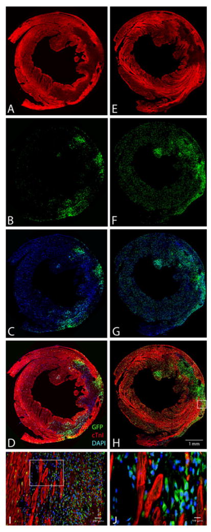
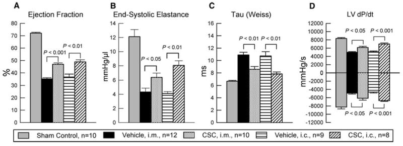
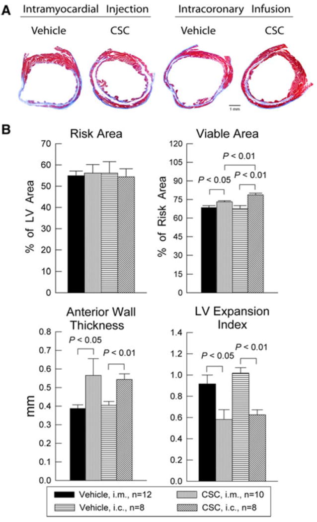
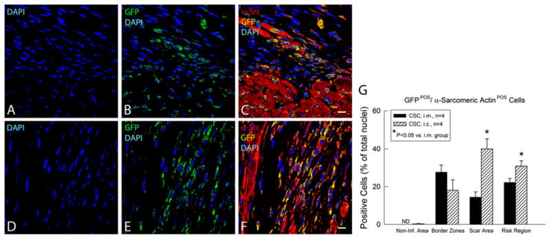
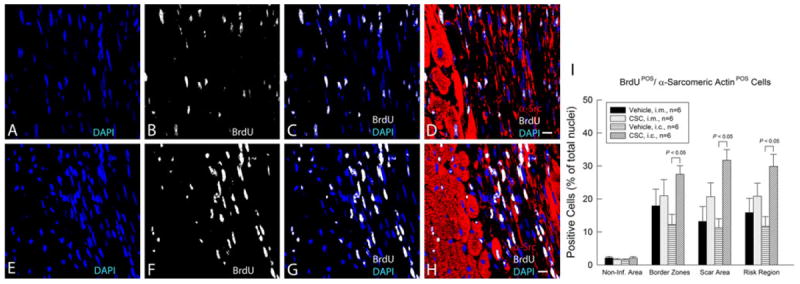
A model of intracoronary stem cell delivery that enables transgenesis/gene targeting would be a powerful tool but is still lacking. To address this gap, we compared intracoronary and intramyocardial delivery of lin(-)/c-kit(+)/GFP(+) cardiac stem cells (CSCs) in a murine model of reperfused myocardial infarction (MI). Lin(-)/c-kit(+)/GFP(+) CSCs were successfully expanded from GFP transgenic hearts and cultured with no detectable phenotypic change for up to ten passages. Intracoronary delivery of CSCs 2 days post-MI resulted in significant alleviation of adverse LV remodeling and dysfunction, which was at least equivalent, if not superior, to that achieved with intramyocardial delivery. Compared with intramyocardial injection, intracoronary infusion was associated with a more homogeneous distribution of CSCs in the infarcted region and a greater increase in viable tissue in this region, suggesting greater formation of new cardiomyocytes. Intracoronary CSC delivery resulted in improved function in the infarcted region, as well as in improved global LV systolic and diastolic function, and in decreased LV dilation and LV expansion index; the magnitude of these effects was similar to that observed after intramyocardial injection. We conclude that, in the murine model of reperfused MI, intracoronary CSC infusion is at least as effective as intramyocardial injection in limiting LV remodeling and improving both regional and global LV function. The intracoronary route appears to be superior in terms of uniformity of cell distribution, myocyte regeneration, and amount of viable tissue in the risk region. To our knowledge, this is the first study to report that intracoronary infusion of stem cells in mice is feasible and effective.


Figures

Fig. 1
Fig. 1
Experimental protocol. Mice were subjected a 60-min coronary occlusion followed by 41 days of reperfusion. CSCs or vehicle were delivered at 48 h after reperfusion. Serial echocardiographic studies were performed at baseline (4 days prior to coronary occlusion), 2 days after CSC transplantation (4 days after reperfusion), and 35 days after CSC transplantation (37 days after reperfusion, and 4 days prior to euthanasia). To assess CSC proliferation, mice were given BrdU (33 mg/kg/day, using mini-osmotic pumps) throughout the 39-day follow-up period after CSC transplantation. At 39 days after CSC transplantation (41 days after reperfusion), mice underwent a hemodynamic study and were euthanized, after which the hearts were harvested for morphometric and histopathologic analyses
Fig. 2
Fig. 2
Isolation and characterization of murine CSCs. At 7 days after primary culture of a fragment of atrial myocardium obtained from a GFPTg mouse heart, GFP+ cells grew around the periphery of the spherical tissue aggregate (a, phase contrast; b, epifluorescence). c Profile of the sorted lin/c-kit+ CSCs (passage 4) labeled with anti-c-kit antibody (red and blue, duplicate assays), labeled with secondary antibody only (black), or unstained (olive). FACS analysis shows that c-kit expression is very high (>95%) and is stable for up to ten passages in cultured CSCs (d), which is consistent with the results of the Western blotting analysis of three independent CSC protein samples (passage 6) (e). f The same three independent CSC protein samples (passage 6) show GFP expression by Western blotting analysis, indicating that these cells are c-kit+/GFP+. g FACS analysis demonstrates robust GFP expression in >99% of the sorted lin/c-kit+/GFP+ CSCs (passage 5). h smears of the sorted lin/c-kit+/GFP+ CSCs expressing c-kit (red) and GFP (green). Nuclei are stained with DAPI (blue). Bars 10 μm
Fig. 3
Fig. 3
Profile and differentiation of murine CSCs. a–c Lin/c-kit+/GFP+ CSCs cultured in growth medium at passage 4 express GFP in the cytoplasm (green) and, in some cases, the early cardiac transcription factor Nkx-2.5 in the nucleus (red). d–f: Lin/c-kit+/GFP+ CSCs cultured in growth medium at passage 2 express c-kit on the membrane (red) and, in some cases, the early cardiac transcription factor MEF2C in the nucleus (green). CSCs cultured in differentiation medium in Matrigel for 10 days (g–h) or in the culture plate without Matrigel for 50 days (i) express the cardiac marker α-sarcomeric actin in the cytoplasm (red) (g–i) and display a cardiac myocyte-like morphology (i). j A 3-D computer reconstruction based on 42 confocal microscopic images showing a CSC that expresses c-kit (red) on the membrane and the early cardiac transcription factor MEF2C (green) in the nucleus. The nuclear membrane is stained with DAPI (blue). k FACS analysis of CSCs cultured for 10 days in differentiation medium shows increased expression of the lineage markers Ets-1 (endothelial), GATA-6 (smooth muscle), and GATA-4, MEF2C, and Nkx-2.5 (cardiac), demonstrating that lin/c-kit+/GFP+ CSCs are heterogeneous and possess the potential to commit to all three components of the cardiac lineage. Data are mean ± SEM. Bars 10 μm
Fig. 4
Fig. 4
Murine CSC migration toward increasing concentrations of SDF-1α. Migration was determined with the Boyden chamber assay. Shown are results obtained from four independent experiments performed in duplicate. Data are mean ± SEM. n = 4/group
Fig. 5
Fig. 5
Distribution of GFP+ CSCs in the heart following transplantation. Six mice underwent a 60-min coronary occlusion followed by reperfusion; CSCs were transplanted 48 h after reperfusion either via intramyocardial injection (a–d) or via intracoronary infusion (e–h). Shown are representative microscopic images of LV sections obtained 2 days after CSC transplantation (i.e., 4 days after reperfusion). Immunofluorescent staining is illustrated for GFP (green), troponin I (red), and DAPI (blue). i is a higher magnification image of the box in h, and j is a higher magnification image of the box in i. These analyses were repeated in three mice that received intramyocardial injection and in three mice that received intracoronary injection
Fig. 6
Fig. 6
Effect of transplantation of CSCs on LV function. Serial echocardiographic studies were performed at baseline (BSL, 4 days prior to coronary occlusion/reperfusion), 48 h after CSC treatment (4 days after reperfusion), and 35 days after CSC transplantation (37 days after reperfusion and 4 days prior to euthanasia). i.m., intramyocardial injection; i.c., intracoronary infusion. Data are mean ± SEM
Fig. 7
Fig. 7
Hemodynamic assessment of LV function at 39 days after intracoronary infusion (i.c.) or intramyocardial injection (i.m.) of CSCs and in age-matched mice not subjected to surgery (sham control group). The figure shows LV ejection fraction, end-systolic elastance, Tau (Weiss), and LV dP/dt. Data are mean ± SEM
Fig. 8
Fig. 8
Morphometric analysis of LV remodeling after CSC transplantation. a Representative Masson’s trichrome-stained myocardial sections from the intramyocardial injection groups (i.m.) treated with vehicle or CSCs, and from the intracoronary infusion groups (i.c.) treated with vehicle or CSCs. Scar tissue and viable myocardium are identified in blue and red, respectively. b quantitative analysis of LV morphometric parameters. LV left ventricle; LV expansion index = (LV cavity area/LV total area) × (non-infarcted region wall thickness/risk region wall thickness). Data are mean ± SEM
Fig. 9
Fig. 9
Fate of transplanted CSCs 39 days after intramyocardial injection (i.m.) or intracoronary infusion (i.c.). Survival and differentiation of transplanted CSCs were determined by immunofluorescent detection of GFP (green), α-sarcomeric actin (red), GFP/α-sarcomeric actin double positivity (yellow), and nuclear DAPI (blue) in hearts that received CSCs either by the i.m. (a–c) or by the i.c (d–f) route. Shown are representative confocal microscopic images obtained from the infarcted area 39 days after CSC transplantation. g Quantitative analysis of the number of GFP/α-sarcomeric actin double positive cells. Data are mean ± SEM. ND Non-Detectable. The region at risk comprises both the border zones and the scar area. Bars 10 μm
Fig. 10
Fig. 10
Proliferation of transplanted CSCs over the 39-day period after intramyocardial injection (i.m.) or intracoronary infusion (i.c.). CSCs were given 48 h after reperfusion by the i.m. or the i.c. route; mice received BrdU beginning at the time of cell transplantation and continuing until euthanasia (for 39 days). Positivity for BrdU (white) in representative confocal microscopic images obtained from the infarcted area identifies newly formed cells (a–d, i.m. group; e–h, i.c. group). α-Sarcomeric actin is red. Nuclei are stained with DAPI (blue). i Quantitative analysis of the number of BrdU/α-sarcomeric actin double positive cells (newly formed cells with apparent commitment to myocytic differentiation) at 39 days after CSC transplantation. Data are mean ± SEM. The region at risk comprises both the border zones and the scar area. Bars 10 μm

References

    1. Abdel-Latif A, Bolli R, Tleyjeh IM, Montori VM, Perin EC, Hornung CA, Zuba-Surma EK, Al-Mallah M, Dawn B. Adult bone marrow-derived cells for cardiac repair: a systematic review and meta-analysis. Arch Intern Med. 2007;167:989–997. doi: 10.1001/archinte.167.10.989. - DOI - PubMed
    1. Anversa P, Kajstura J, Leri A. If I can stop one heart from breaking. Circulation. 2007;115:829–832. doi: 10.1161/CIRCULATIONAHA.106.682195. - DOI - PubMed
    1. Bearzi C, Rota M, Hosoda T, Tillmanns J, Nascimbene A, De Angelis A, Yasuzawa-Amano S, Trofimova I, Siggins RW, Lecapitaine N, Cascapera S, Beltrami AP, D’Alessandro DA, Zias E, Quaini F, Urbanek K, Michler RE, Bolli R, Kajstura J, Leri A, Anversa P. Human cardiac stem cells. Proc Natl Acad Sci USA. 2007;104:14068–14073. doi: 10.1073/pnas.0706760104. - DOI - PMC - PubMed
    1. Beltrami AP, Barlucchi L, Torella D, Baker M, Limana F, Chimenti S, Kasahara H, Rota M, Musso E, Urbanek K, Leri A, Kajstura J, Nadal-Ginard B, Anversa P. Adult cardiac stem cells are multipotent and support myocardial regeneration. Cell. 2003;114:763–776. - PubMed
    1. Black RG, Jr, Guo Y, Ge ZD, Murphree SS, Prabhu SD, Jones WK, Bolli R, Auchampach JA. Gene dosage-dependent effects of cardiac-specific overexpression of the A3 adenosine receptor. Circ Res. 2002;91:165–172. - PMC - PubMed

Publication types

MeSH terms

Substances