. 2011 Jun;117(6):1033-46.
doi: 10.1111/j.1471-4159.2011.07279.x. Epub 2011 May 13.

Nucleolar disruption and apoptosis are distinct neuronal responses to etoposide-induced DNA damage

Affiliations

Nucleolar disruption and apoptosis are distinct neuronal responses to etoposide-induced DNA damage

Maciej Pietrzak et al. J Neurochem. 2011 Jun.

Abstract

Although DNA damaging topoisomerase inhibitors induce apoptosis in developing neurons, their effects on adult neurons have not yet been characterized. We report a blockage of RNA-Polymerase-1-driven transcription and nucleolar stress in neocortical neurons of adult rats after intracarotid injection of the DNA-topoisomerase-2 inhibitor, etoposide. Intracerebroventricular injection of etoposide induced a similar response in neonatal rats. In contrast, etoposide triggered neuronal apoptosis in the neonates, but not the adults. Nucleolar disruption and apoptosis were also observed in etoposide-challenged cultured cortical neurons from newborn rats. In that system, activation of the DNA double strand break signaling kinase ataxia telangiectasia-mutated protein kinase, p53 and p53-dependent apoptosis required lower etoposide concentrations than did the p53-independent induction of nucleolar stress. These distinct responses may be coupled to different forms of etoposide-induced DNA damage. Indeed, double strand breaks by the over-expressed endonuclease I-Ppo1 were sufficient to induce p53-dependent apoptosis. Moreover, nucleolar transcription was insensitive to such damage implying single strand breaks and/or topoisomerase-2-DNA adducts as triggers of nucleolar stress. Because nucleolar stress is not age-restricted, it may underlie non-apoptotic neurotoxicity of chemotherapy- or neurodegeneration-associated DNA damage by reducing ribosomal biogenesis in adult brain. Conversely, nucleolar insensitivity to double strand breaks likely contributes to mature neuron tolerance of such lesions.


Figures

Figure 1
Figure 1. Nucleolar disruption in the neocortex of etoposide-treated adult rats
Rats were infused with 0 or 15 mg/kg etoposide that was administered into the right intracarotid artery immediately after BBB disruption with 20% d-mannitol. A, At 4 h after etoposide treatment, reduced nucleolar transcription was observed in the ipsilateral neocortex as indicated by the declining expression ratio of the primary nucleolar rRNA transcript (45S pre-rRNA) to the mature 18S rRNA. The rRNA levels were determined by quantitative real-time PCR; data are means ±SE of 4 animals per group; *, p<0.05. B, At 6 h after etoposide treatment, nucleolar stress was observed throughout the ipsilateral neocortex as indicated by the loss of nucleolar immunofluorescence of nucleolphosmin/B23. B23, which under control conditions was concentrated in nucleoli (arrows), displayed a diffused staining pattern throughout the nucleoplasm in etoposide-infused rats (arrowheads). Note that cells with nucleolar stress had normal chromatin structure as visualized by counterstaining with Hoechst-33258. Representative photomicrographs of cortical layer IV-VI are shown; similar effects of etoposide on B23 localization were observed in 3 animals.
Figure 2
Figure 2. Nucleolar disruption in the neocortex of etoposide-treated newborn rats
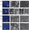
At postnatal day 7 (P7), rats received 0 or 10 nmoles etoposide that was administered by an injection into the right lateral ventricle. A, Four hours after etoposide injection, nucleolar transcription was suppressed in the ipsilateral neocortex. Data are means ±SE of 4 animals per group; **, p<0.01. B, Six hours after etoposide injection, nucleolar stress was present in the ipsilateral neocortex. Reduced nucleolar B23 staining together with its diffused appearance throughout the nucleoplasm was observed in most neocortical cells (arrowheads). B23 under control conditions was concentrated in nucleoli (arrows). Note that cells with nucleolar stress had normal chromatin structure as visualized by counterstaining with Hoechst-33258. Representative photomicrographs of cortical layer IV-VI are shown; similar effects of etoposide on B23 localization were observed in 4 animals.
Figure 3
Figure 3. Etoposide-induced apoptosis in the neocortex of the neonate, but not the adult rats
Neonate (P7) (A-H) or adult rats (I-N) were treated as described for Figs. 2, and 1, respectively. Six hours after etoposide (Etop) administration apoptosis was identified by the presence of immunofluorescence for activated caspase-3 and/or condensation/fragmentation of nuclear chromatin that was visualized by counterstaining with Hoechst-33258. Photomicrographs of the ipsilateral neocortex are shown. Three animals were analyzed for each condition. Cortical layers are indicated by roman numerals. A-H, Activated caspase-3 and apoptotic chromatin rearrangements in neocortical cells of etoposide-treated neonates (arrows). Presence of active caspase-3 in neurites suggests neuronal identity of many apoptotic cells (arrowheads). I-N, Absence of apoptotic cells from the neocortex of etoposide-treated adult rats.
Figure 4
Figure 4. Etoposide-induced apoptosis in the hippocampus of the neonate, but not the adult rats
Neonate (P7) (A-F) or adult rats (G-L) were treated as described for Figs. 2, and 1, respectively. Six hours after etoposide (Etop) administration apoptosis was identified as in Fig. 3. Photomicrographs of the ipsilateral hippocampi are shown. Three animals were analyzed for each condition. A-F, Etoposide-induced activation of caspase-3, and apoptotic chromatin rearrangements in granule cells of the neonate dentate gyrus (arrows). G-L, Absence of apoptotic cells from the dentate gyrus of etoposide-treated adult rats.
Figure 5
Figure 5. Etoposide-induced nucleolar stress in cultured cortical neurons from newborn rats
At DIV5, rat cortical neurons were treated with etoposide for 8 h as indicated. A, At etoposide concentrations of 25- or 50- but not 10 μM, the ratios of 45S pre-rRNA/18S rRNA levels declined suggesting inhibition of Pol1-mediated transcription. B, At etoposide concentrations of 25- or 50- but not 10 μM, B23 immunofluorescence was present not only in the nucleoli, but also throughout the nucleolplasm indicating nucleolar stress. However, even at 50 μM, etoposide did not completely disrupt nucleolar B23 staining. In contrast, no nucleolar B23 was observed in neurons treated with the Topo1 inhibitor camptothecin (5 μM, CPT) that was used as a positive control. Note that after an 8 h etoposide treatment, neurons with nucleoplasmic translocation of B23 maintained normal structure of nuclear chromatin as indicated by Hoechst-33258 staining. C-D, Quantitative analysis of B23 immunofluorescence after treatment with 50 μM etoposide. Etoposide reduced nucleolar intensity of B23 immunofluorescence (C) while increasing it in the nucleoplasm (D). In A, C, and D, data represent means ±SE of three independent experiments; in C, and D, 10 randomly selected cells were analyzed for each experiment; *, p<0.05; **, p<0.01; ***, p<0.001. Dose response trends similar to those shown in B were observed in four independent experiments.
Figure 6
Figure 6. Apoptosis of cultured cortical neurons at lower etoposide concentrations than nucleolar stress
At DIV5, rat cortical neurons were treated with etoposide as indicated. A, After 8 h incubation with etoposide at such low concentrations as 2.5 μM, nearly maximal activation of caspase-3 was revealed by immunoblotting. Equal protein loading was ensured by re-probing forβ-actin. B, After a 24 h etoposide treatment, Hoechst-33258 staining uncovered apoptotic changes in nuclear chromatin including its uniform condensation with or without fragmentation. C, Dose response analysis of etoposide-induced apoptotic chromatin condensation indicated that at 10 μM, etoposide triggered maximum apoptosis. Data represent means ±SE of three independent experiments.
Figure 7
Figure 7. Role of the p53 pathway in etoposide-induced apoptosis but not nucleolar stress
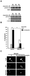
DIV5 cortical neurons were treated with etoposide as indicated. A, Immunoblot analysis of the DNA damage-induced autophosphorylation of the protein kinase ATM at the Ser-1981 residue (pS1981-ATM) and activation-associated phosphorylation of the ATM downstream target p53 (phospho-Ser15, pS15-p53). In addition, total levels of p53 were also determined. Equal protein loading of the blots was ensured by reprobing for MAP2 or β-actin as indicated. At the pro-apoptotic concentration of 10 μM, etoposide increased pS1981-ATM, pS15-p53, and total p53 levels suggesting activation of the ATM/p53 signaling. B, At DIV4, cortical neurons were co-transfected with the expression plasmids for dominant-negative p53 (DN-p53) and β-gal (EF1αLacZ, 0.2+0.2 μg/5 × 105 cells, respectively). The empty vector pcDNA3.1(+) was used as a negative control (vector). Etoposide was added 24 h post-transfection. After a 24 h treatment, transfected cells were detected by β-gal immunofluorescence and apoptotic chromatin rearrangements were visualized by counterstaining with Hoechst-33258. C, Cells were transfected as in B. After an 8 h treatment with etoposide B23 immunofluorescence was analyzed. Transfected cells were detected by co-staining against β-gal (arrows). Despite inhibition of the p53 pathway, nucleolar stress was present in etoposide-treated neurons as indicated by the diffused nucleolplasmic appearance of B23 (arrowheads). In A, and C, similar trends were observed in three independent experiments; in B, means ±SE of three independent experiments are depicted; **, p<0.01; ***, p<0.001.
Figure 8
Figure 8. Lack of nucleolar stress or apoptosis in cultured neurons treated with ICRF-193, the non-DNA-damaging inhibitor of DNA-topoisomerase-2
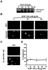
At DIV5, cortical neurons were treated with ICRF-193 as indicated. A, Immunoblot analysis of the major neuronal isoform of Topo2, Topo2β. ICRF-193 induced shifts in its electromobility that are consistent with inhibition-associated ubiquitination of Topo2β. B-D, ICRF-193 affected neither nucleolar B23 immunofluorescence (B) nor frequency of apoptotic cells (C-D) suggesting that the non-genotoxic inhibition of Topo2 is insufficient to induce nucleolar stress or apoptosis. In C, representative photomicrographs of Hoechst-33258-stained cells are shown; note that ICRF-193-treated neurons display normal non-apoptotic chromatin. Similar trends to those in A, and, B were observed in two independent experiments; in D, means ±SE of three independent experiments are depicted.
Figure 9
Figure 9. The selective DSB DNA damage is sufficient to induce the p53-dependent apoptosis of cultured cortical neurons
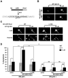
A, The homing endonuclease I-Ppo1 was overexpressed as a fusion protein with a hemaglutinin (HA) epitope tag and a mutant ligand binding domain of the estrogen receptor (mER). Hence, its nuclear translocation is stimulated by the mER ligand 4-hydroxytamoxifen (4HT). In the nucleus, I-Ppo1 induces a DSB within a 15 bp site in the 28S portion of the rDNA and also at some extranucleolar locations (Table 1). The nuclease-dead mutant variant of I-Ppo1 (L116A) can not induce DSBs. B, At DIV4, cortical neurons were co-transfected with the expression plasmids for β-gal (EF1αlacZ) and the wt- or the L116A mutant I-Ppo1 (pRc/RSV-HA-mER-I-Ppo1wt/L116A; 0.2+1 μg plasmid DNA/5 × 105 cells, respectively). The empty cloning vector pRc/RSV (Vector) was used as an additional control. Four-HT was added 24 h post-transfection as indicated. After an 8 h 4HT treatment, the fusion protein HA-mER-I-Ppo1 accumulated in the nucleus as visualized by anti-HA immunostaining (arrowheads). C, After a 24 h 4HT treatment, wt- but not L116A I-Ppo1 induced neuronal apoptosis. Transfected cells were detected by β-gal immunofluorescence, apoptotic changes in nuclear morphology were visualized by counterstaining with Hoechst-33258. Arrows indicate transfected cells, an apoptotic cell is pointed by an arrowhead. D, Neurons were transfected as in B. In addition, DN-p53 expression construct (DN-p53+) or an empty cloning vector control (pcDNA3.1, DN-p53-) was added to the transfections (0.2 μg plasmid DNA/5 × 105 cells). The 4HT treatment and apoptosis analysis were as in C. 4HT increased apoptosis in neurons transfected with I-Ppo1wt, but not L116A. That effect was prevented by the DN-p53. Means ±SE of three independent experiments are depicted; ns, p>0.05; *, p<0.05; ***, p<0.001.
Figure 10
Figure 10. Insufficiency of the selective DSB DNA damage to induce nucleolar stress in cultured cortical neurons
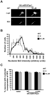
Cortical neurons were transfected as in Fig. 9D. DN-p53 was added to all transfections to prevent I-Ppo1-induced apoptosis. A-B, The activated I-Ppo1 did not disrupt nucleolar B23 immunofluorescence. In transfected neurons that were detected by the positive β-gal immunostaining (arrows), 4HT induced neither nucleoplasmic translocation of B23 (A) nor its disappearance from the nucleoli (B). C, Nucleolar transcription is unaffected by the activated I-Ppo1. In situ run-on assay with the RNA precursor 5FU was used to monitor nucleolar transcription in transfected neurons that overexpressed wt- or L116A I-PPo1. The fraction of cells with nucleolar accumulation of nascent RNA was similar regardless of the transfected plasmid or the 4HT treatment. In B, and C, data are means ±SD of two independent experiments.

Similar articles

Cited by

References

    1. Berkovich E, Monnat RJ, Jr, Kastan MB. Roles of ATM and NBS1 in chromatin structure modulation and DNA double-strand break repair. Nat Cell Biol. 2007;9:683–690. - PubMed
    1. Brasnjevic I, Hof PR, Steinbusch HW, Schmitz C. Accumulation of nuclear DNA damage or neuron loss: molecular basis for a new approach to understanding selective neuronal vulnerability in neurodegenerative diseases. DNA Repair (Amst) 2008;7:1087–1097. - PMC - PubMed
    1. Brooks PJ. The 8,5′-cyclopurine-2′-deoxynucleosides: candidate neurodegenerative DNA lesions in xeroderma pigmentosum, and unique probes of transcription and nucleotide excision repair. DNA Repair (Amst) 2008;7:1168–1179. - PMC - PubMed
    1. Burger K, Muhl B, Harasim T, et al. Chemotherapeutic drugs inhibit ribosome biogenesis at various levels. J Biol Chem. 2010;285:12416–12425. - PMC - PubMed
    1. Caldecott KW. Single-strand break repair and genetic disease. Nat Rev Genet. 2008;9:619–631. - PubMed

Publication types

MeSH terms