. 2011 May-Jun;19(3):426-35.
doi: 10.1111/j.1524-475X.2011.00682.x. Epub 2011 Apr 21.

The influence of interleukin-4 on ligament healing

Affiliations

The influence of interleukin-4 on ligament healing

Connie S Chamberlain et al. Wound Repair Regen. 2011 May-Jun.

Abstract

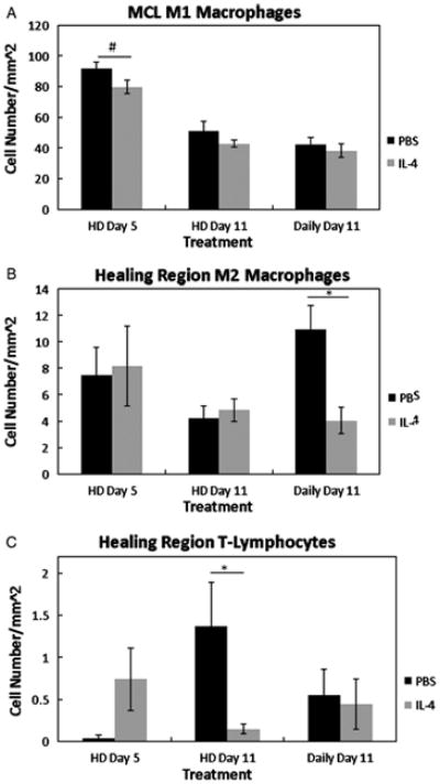
Despite a complex cascade of cellular events to reconstruct the damaged extracellular matrix, ligament healing results in a mechanically inferior scarred ligament. During normal healing, granulation tissue expands into any residual normal ligamentous tissue (creeping substitution), resulting in a larger region of healing, greater mechanical compromise and an inefficient repair process. To control creeping substitution and possibly enhance the repair process, the antiinflammatory cytokine, interleukin-4 (IL-4), was administered to rats before and after rupture of their medial collateral ligaments. In vitro experiments showed a time-dependent effect on fibroblast proliferation after IL-4 treatment. In vivo treatments with IL-4 (100 ng/mL IV) for 5 days resulted in decreased wound size and type III collagen and increased type I procollagen, indicating a more regenerative early healing in response to the IL-4 treatment. However, continued treatment of IL-4 to day 11 antagonized this early benefit and slowed healing. Together, these results suggest that IL-4 not only influences the macrophages and T lymphocytes but also stimulates fibroblasts associated with the proliferative phase of healing in a dose-, cell-, and time-dependent manner. Although treatment significantly influenced healing in the first week after injury, IL-4 alone was unable to maintain this early regenerative response.


Figures

Figure 1
Figure 1
Representative cross-section of an H&E-stained healing MCL, indicating the approximate locations subsequent images were captured for later cell enumeration. Two to three sections from each animal were examined accordingly (HR, healing region; pHR, proximal healing region; dHR, distal healing region; prox, proximal; EL, epiligament; Original magnification 40X).
Figure 2
Figure 2
Dose response of IL-4 on fibroblast proliferation from cells obtained from uninjured (A) or day 5 post-injured ligaments (B). Cells were collected at 24, 48 and 72 (not shown) hours post-treatment and quantified.
Figure 3
Figure 3
Graph of granulation tissue size after IL-4 treatment (n=3 rats/treatment; 21 total rats). High doses of IL-4 significantly reduced the normalized healing region at day 5 (p < .05). No other significant treatment effects were observed at day 11 or with low-doses of IL-4. Values are the mean percentage of the granulation tissue area divided by the total ligament area ± S.E.M.
Figure 4
Figure 4
Graph of the M1 macrophages (A), M2 macrophages (B), and T-lymphocytes (C), at 5 and 11 days post-injury after PBS or IL-4 treatment (n=3 rats/treatment; 21 total rats). On day 5, high doses of IL-4 (HD Day 5) tended to reduce the MCL M1 macrophages (p =.078; A). No differences within the MCL were observed at any other points. The day 11 healing region M2 macrophages were significantly reduced after continuous treatment of IL-4 (Daily Day 11;B). High doses of IL-4 (HD Day 5) reduced the number of healing region T-lymphocytes at day 11.#Indicates a trend (p < .1 between PBS and IL-4 at day 5. * Indicates significance (p< .05) between PBS and IL-4 at day 11 (C). Values are expressed as mean cell numbers ± S.E.M.
Figure 5
Figure 5
Graph of type I procollagen (A) and type III collagen (B) after PBS and IL-4 treatment (n=3 rats/treatment; 21 total rats). High doses of IL-4 (HD Day 5) significantly increased type I procollagen (A) and decreased type III collagen (B) at day 5 within the MCL. However, daily doses of IL-4 (Daily Day 11) reduced type I collagen (p= .057; A). No other effects were observed with type III collage at day 11 (B).*Indicates significant difference (p < .05) between PBS and IL-4 at day 5 (A, B) or day 11 (A). #Indicates a trend (p < 0.1 between PBS and IL-4 at day 11 (Daily Day 11). Values are expressed as mean positive staining ± S.E.M.
Figure 6
Figure 6
Representative micrograph of H&E (A–B), Type I procollagen (C–F) and Type III collagen (G–H) after PBS (left column) or IL-4 (right side) treatment. Size of the day 5 granulation tissue after PBS (A) or IL-4 (B) treatment. Black circles are the same size to compare the granulation size differences between the two groups (A–B). Type I procollagen IHC of the day 5 ligament after treatment with PBS (C) or high doses of IL-4 (D). Type I procollagen IHC of the day 11 ligament after PBS treatment (E) or daily high doses of IL-4 (F). Type III collagen IHC of the day 5 ligament after PBS (G) or high doses of IL-4 (H) treatment.
Figure 7
Figure 7
Response of endothelial cells (A) and myofibroblasts (B) to PBS or IL-4 (n=3 rats/treatment; 21 total rats). No treatment effects were observed at day 5 (A, B). In contrast IL-4 significantly reduced the number of MCL endothelial cells at day 11 (A). Healing region myofibroblasts were also reduced with daily doses of IL-4 at day 11 whereas injections up to day 4 were not effective (B). *Indicates significant difference (p < .05) between PBS and IL-4 at day 11 (A, B). Values are expressed as mean positive staining ± S.E.M.
Figure 8
Figure 8
Failure force (A), failure stress (B), and stiffness (C) to response to PBS or IL-4 treatment 11 days post-injury(n=5 rats/treatment; 10 total rats). No significance in failure force (A) failure stress (B) or stiffness (C) was observed. P > .05. Results are expressed as mean ± S.E.M.

References

    1. Clark RA, Nielsen LD, Welch MP, McPherson JM. Collagen matrices attenuate the collagen-synthetic response of cultured fibroblasts to TGF-beta. Journal of cell science. 1995;108 (Pt 3):1251–1261. - PubMed
    1. Singer AJ, Clark RA. Cutaneous wound healing. The New England journal of medicine. 1999;341:738–746. - PubMed
    1. Levenson SM, Geever EF, Crowley LV, Oates JF, 3rd, Berard CW, Rosen H. The healing of rat skin wounds. Annals of Surgery. 1965;161:293–308. - PMC - PubMed
    1. Lin TW, Cardenas L, Soslowsky LJ. Biomechanics of tendon injury and repair. Journal of Biomechanics. 2004;37:865–877. - PubMed
    1. Schnabel LV, Lynch ME, van der Meulen MC, Yeager AE, Kornatowski MA, Nixon AJ. Mesenchymal stem cells and insulin-like growth factor-I gene-enhanced mesenchymal stem cells improve structural aspects of healing in equine flexor digitorum superficialis tendons. Journal of orthopaedic research: official publication of the Orthopaedic Research Society. 2009;27:1392–1398. - PubMed

Publication types

MeSH terms