. 2011 Jun 10;286(23):20547-57.
doi: 10.1074/jbc.M111.230151. Epub 2011 Apr 25.

Tapasin discriminates peptide-human leukocyte antigen-A*02:01 complexes formed with natural ligands

Affiliations

Tapasin discriminates peptide-human leukocyte antigen-A*02:01 complexes formed with natural ligands

Gustav Roder et al. J Biol Chem. .

Abstract

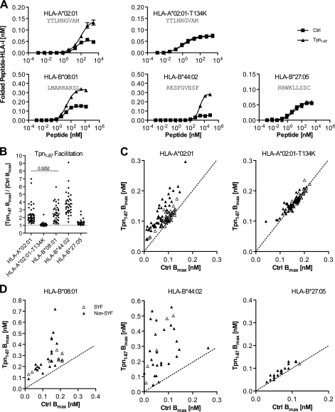
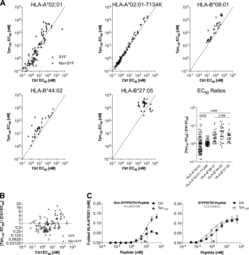
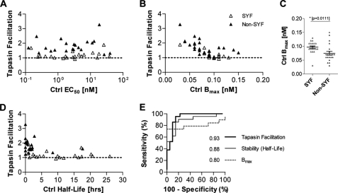
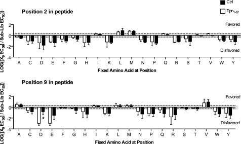
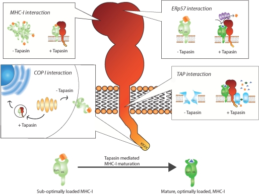
A plethora of peptides are generated intracellularly, and most peptide-human leukocyte antigen (HLA)-I interactions are of a transient, unproductive nature. Without a quality control mechanism, the HLA-I system would be stressed by futile attempts to present peptides not sufficient for the stable peptide-HLA-I complex formation required for long term presentation. Tapasin is thought to be central to this essential quality control, but the underlying mechanisms remain unknown. Here, we report that the N-terminal region of tapasin, Tpn(1-87), assisted folding of peptide-HLA-A*02:01 complexes according to the identity of the peptide. The facilitation was also specific for the identity of the HLA-I heavy chain, where it correlated to established tapasin dependence hierarchies. Two large sets of HLA-A*02:01 binding peptides, one extracted from natural HLA-I ligands from the SYFPEITHI database and one consisting of medium to high affinity non-SYFPEITHI ligands, were studied in the context of HLA-A*02:01 binding and stability. We show that the SYFPEITHI peptides induced more stable HLA-A*02:01 molecules than the other ligands, although affinities were similar. Remarkably, Tpn(1-87) could functionally discriminate the selected SYFPEITHI peptides from the other peptide binders with high sensitivity and specificity. We suggest that this HLA-I- and peptide-specific function, together with the functions exerted by the more C-terminal parts of tapasin, are major features of tapasin-mediated HLA-I quality control. These findings are important for understanding the biogenesis of HLA-I molecules, the selection of presented T-cell epitopes, and the identification of immunogenic targets in both basic research and vaccine design.


Figures

FIGURE 1.
FIGURE 1.
Tpn1–87 facilitates folding of peptide-HLA-I complexes according to peptide and HLA-I identity. A, folding of peptide-HLA-I complexes in single peptide dose-response experiments. Fixed concentrations of β2m and HLA-I HCs were mixed with titrated concentrations of peptide in the presence (▴) or absence (■) of Tpn1–87. The mixtures were incubated at 18 °C for 48 h, and folded peptide-HLA-I complexes were detected by the HLA-I conformation-specific W6/32 monoclonal antibody in a homogenous assay (36). B, a study of Tpn1–87 facilitation based on the maximum amount of folded peptide-HLA-I complexes, Bmax. Peptide dose-response curves were made by offering each peptide in different concentrations to the folding reaction. The saturation plateaus were calculated as Bmax from the curves. Binding curves were made in the presence (Tpn1–87 Bmax) and absence (Ctrl Bmax) of Tpn1–87 with SYFPEITHI (△) and non-SYFPEITHI (▴) peptides. B, the degree of Tpn1–87 facilitation for each of the tested HLA-I molecules is shown. C, the Bmax values with and without Tpn1–87 for the binding of each of the tested peptides to HLA-A*02:01 and HLA-A*02:01-T134K are plotted. D, the Bmax values for the binding of peptides specific for HLA-B*44:02, -B*08:01, and -B*27:05 with and without Tpn1–87 are plotted. All of the experiments were done in quadruplicate, and standard deviations for each folding reaction were calculated and visualized in the graphs. A Student's t test was applied to determine whether the means were significantly different. All of the means were significantly different (p < 0.001), except for the one with the p value shown in the graph.
FIGURE 2.
FIGURE 2.
Tpn1–87 influences the peptide binding affinity to HLA-I. A, Tpn1–87 influences the peptide affinity to HLA-I. Multiple binding curves were made, and the peptide concentration resulting in half-saturation was calculated as EC50. Binding curves were made in the presence (Tpn1–87 EC50) and absence (Ctrl EC50) of Tpn1–87. 44 SYFPEITHI (△) and 44 non-SYFPEITHI (▴) peptides were tested on HLA-A*02:01. The same peptides were tested on HLA-A*02:01-T134K. Other peptide panels were tested on HLA-B*08:01, HLA-B*44:02, and HLA-B*27:05. The EC50 ratios (Tpn1–87 EC50/Ctrl EC50) are shown in the EC50 ratios graph. A Student's t test was used to determine whether the means were significantly different (p < 0.05) between the HLA-I molecules. The p values are shown in cases where no significant differences were found (for a complete list see supplemental Table S2). B, the EC50 ratios on HLA-A*02:01 were plotted against Ctrl EC50 and grouped in non-SYFPEITHI and SYFPEITHI peptide groups. C, peptide dose-response curves representing the non-SYFPEITHI and SYFPEITHI peptide-HLA-A*02:01 complexes were analyzed for shifts in EC50. For the non-SYFPEITHI peptide, the EC50 values increased in the presence of Tpn1–87 corresponding to a decrease in affinity. For the SYFPEITHI peptide, the EC50 values decreased in the presence of Tpn1–87, corresponding to an increase in affinity.
FIGURE 3.
FIGURE 3.
Tpn1–87 facilitates folding and discriminates immunogenic peptides independent of peptide affinity to HLA-A*02:01. 21 SYFPEITHI and 21 non-SYFPEITHI peptides were paired, based upon affinity to HLA-A*02:01. Fixed concentrations of β2m and HLA-A*02:01 HC were mixed with various concentrations of peptide in the presence or absence of Tpn1–87. A, the peptide affinities (EC50) to the HLA-I molecules were calculated as the peptide concentration required to reach the half-saturation point on the sigmoidal dose-response curve. The Tpn1–87 facilitation was plotted against EC50. B, the Tpn1–87 facilitation was plotted against the saturation plateau, Bmax. C, the Bmax values for the SYFPEITHI and non-SYFPEITHI peptides in the absence of Tpn1–87 were plotted in a vertical scatter diagram. D, the Tpn1–87 facilitation was plotted against measured stabilities of the peptide-HLA-A*02:01. E, ROC analysis was performed for the ability of each parameter (Bmax, stability, and Tpn1–87 facilitation) to discriminate between SYFPEITHI peptides and non-SYFPEITHI peptides. The AUC values are shown. To determine whether significant differences exist between the areas under the ROC curves, a jack knife analysis was performed on the ROC areas. A Student's t test was used to determine statistically significant differences (p < 0.05) between the parameters tested. All of the AUCs differed significantly in the t test. ***, p < 0.0001.
FIGURE 4.
FIGURE 4.
Tpn1–87 alters the peptide binding specificity of HLA-A*02:01 to a minor extent. The peptide binding specificity of HLA-A*02:01 was tested using PSCPLs and shown for substitution positions 2 and 9 in the peptide. Log values of RB values (X9 EC50/sublibrary EC50) are plotted on the y axis, and the amino acid substitutions are shown on the x axis. Amino acid substitutions leading to RB values above 0.3 (log(2)) are considered favored, and values below −0.3 (log(0.5)) are considered unfavored (these boundaries are indicated by the gray shading). Significant differences (p < 0.05) are marked with an asterisk.
FIGURE 5.
FIGURE 5.
Different sites and regions of tapasin work in concert to quality control MHC-I. Top left box, entire regions and single residues in tapasin, from the cytoplasmic tail to the most N-terminal region, have been suggested to be involved in MHC-I binding. Tapasin incorporates MHC-I into the PLC and brings it into close proximity of the TAP transported peptides. Major MHC-I binding sites are located in the ER luminal part of tapasin. In the first 87 amino acids of tapasin, a chaperone function is located that is suggested to keep the MHC-I in a peptide-receptive state and prevent MHC-I aggregation and degradation. Top right box, Cys-95 in tapasin forms a disulfide conjugate with Cys-57 in ERp57, which was suggested to allow tapasin to function as a MHC-I peptide editor. Bottom right box, sites in the cytosolic and transmembrane region of tapasin are important for binding to TAP1 and TAP2. Tapasin both stabilizes TAP and promotes binding of peptides to TAP before the ATP-dependent peptide translocation across the ER membrane. Bottom left box, a double lysine motif is located in the C-terminal of tapasin and mediates interaction with coat protein type I (COP I) vesicles. Coat protein type I vesicles have been proposed to recycle immature/peptide-receptive MHC-I molecules from the Golgi back to the ER. Binding of optimal peptide releases MHC-I from tapasin, allowing efficient antigen presentation on the cell surface.

References

    1. Cresswell P., Bangia N., Dick T., Diedrich G. (1999) Immunol. Rev. 172, 21–28 - PubMed
    1. Park B., Lee S., Kim E., Cho K., Riddell S. R., Cho S., Ahn K. (2006) Cell 127, 369–382 - PubMed
    1. Li S., Sjögren H. O., Hellman U., Pettersson R. F., Wang P. (1997) Proc. Natl. Acad. Sci. U.S.A. 94, 8708–8713 - PMC - PubMed
    1. Ortmann B., Copeman J., Lehner P. J., Sadasivan B., Herberg J. A., Grandea A. G., Riddell S. R., Tampé R., Spies T., Trowsdale J., Cresswell P. (1997) Science 277, 1306–1309 - PubMed
    1. Grandea A. G., 3rd, Lehner P. J., Cresswell P., Spies T. (1997) Immunogenetics 46, 477–483 - PubMed

Publication types

MeSH terms