. 2011 May 2;193(3):455-64.
doi: 10.1083/jcb.201011018. Epub 2011 Apr 25.

Cell wound repair in Drosophila occurs through three distinct phases of membrane and cytoskeletal remodeling

Affiliations

Cell wound repair in Drosophila occurs through three distinct phases of membrane and cytoskeletal remodeling

Maria Teresa Abreu-Blanco et al. J Cell Biol. .

Abstract

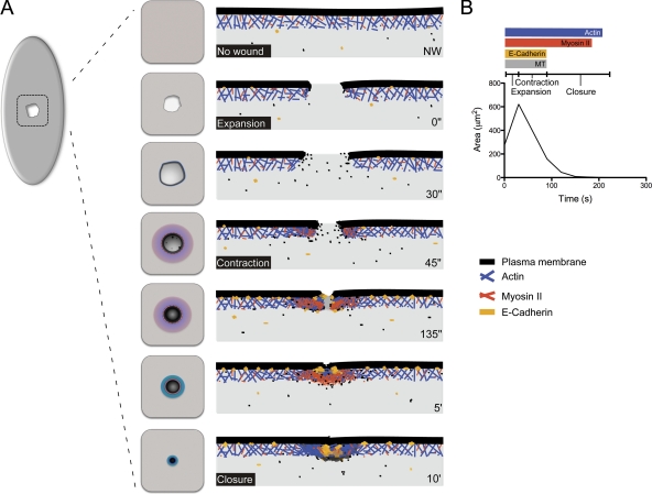
When single cells or tissues are injured, the wound must be repaired quickly in order to prevent cell death, loss of tissue integrity, and invasion by microorganisms. We describe Drosophila as a genetically tractable model to dissect the mechanisms of single-cell wound repair. By analyzing the expression and the effects of perturbations of actin, myosin, microtubules, E-cadherin, and the plasma membrane, we define three distinct phases in the repair process-expansion, contraction, and closure-and identify specific components required during each phase. Specifically, plasma membrane mobilization and assembly of a contractile actomyosin ring are required for this process. In addition, E-cadherin accumulates at the wound edge, and wound expansion is excessive in E-cadherin mutants, suggesting a role for E-cadherin in anchoring the actomyosin ring to the plasma membrane. Our results show that single-cell wound repair requires specific spatial and temporal cytoskeleton responses with distinct components and mechanisms required at different stages of the process.


Figures

Figure 1.
Figure 1.
The Drosophila embryo is a model to study single-cell wound healing. Surface projections (A) and orthogonal sections (C) of early embryos expressing actin and histone (sGMCA; His2Av-mRFP). Nuclear cycle is indicated. (B) Cartoon depicting the embryo stages shown in A. (D and D′) Actin accumulates at the cell wound edge (arrows). Time-lapse series of surface projections (D) and cross sections (D′) of embryos expressing actin (sGMCA). Dotted line in D indicates the plane of the cross section. (E) Analysis of the phases of single cell wound repair (n = 14; results are given as means ± SEM). (F) Effects of wound size in cell wound repair. (small, n = 16; medium, n = 14; large, n = 7). (G–H) Confocal images of wound repair in embryos expressing actin and plasma membrane markers (sChMCA; GFP-Spider) and treated with Lat B (n = 6). Left panel shows actin depolymerization before wounding (circle indicates the ablation site). (G′) Kymograph showing failure of actin cable assembly (W, wound). (G′′) Actin and membrane recruitment are impaired. (H) In embryos exhibiting partial actin depolymerization, the actin cable is assembled in regions of the wound edge richer in actin. (I and I′) Image of a syncytial embryo expressing GFP–α-tubulin. (J and J′) Time series after wounding in embryos treated with colchicine (sGMCA). (J′) Kymograph analysis. (J′′) Ring width quantification (control = 8, colch = 8; P = 0.0003). Bars: (A, D, G, H, I, and J) 20 µm; (C, D′, G′, and I′) 10 µm; (G′′ and J′) 5 µm.
Figure 2.
Figure 2.
Contribution of myosin II to single-cell wound repair. (A–B) Images from early embryos expressing sChMCA; sqh-GFP. (A) Colocalization of myosin II (A′) and actin (A′′). (A′′′) Cross sections showing actin (red arrowhead) and myosin dynamics (green arrowheads). (B) Single Z sections of embryo in A. (C and C′) Confocal time series after the healing process in sqh1 embryos expressing actin (sGMCA; n = 4). (C′) Kymograph analysis, two sqh1 and a control embryo are shown. Top panel corresponds to embryo in C. Bars: (A–C) 20 µm; (A′′′) 10 µm; (C′) 5 µm.
Figure 3.
Figure 3.
Plasma membrane component involvement in single-cell wound repair. Time series of confocal images of early embryos expressing: sChMCA; GFP-Spider (A–A′′), GFP-Spider (B and B′), PH-PLC-GFP (D and D′), and GAP43-ChFP (E–E′′). (A and A′) Actin (red arrowhead) and plasma membrane (green arrowhead) localization at the wound is indicated. (A′′) Single confocal section of specific time points from A, the position of the Z slice is indicated. Fluorescence intensity at the wound edge in SChMCA; GFP-Spider (A′′′), GFP-Spider (C), PH-PLC-GFP (D′′), and GAP43-ChFP (E′′) embryos at specific times after wounding. (F) Cartoon showing the Z planes. Plasma membrane (black line), actin (blue), and myosin (orange) are depicted. Bars: (A, A′′, B, D, E, and E′′) 20 µm; (A′, B′, D′, E′, and E′′′) 10 µm.
Figure 4.
Figure 4.
DE-cadherin is required for cell wound repair. (A–A′′) DE-cadherin was detected by antibody staining in embryos expressing a nuclear marker (His2Av-mRFP). (A′) Single confocal slices of the embryos in A. (B–B′′) Time-lapse series of an early embryo expressing sChMCA; cadherin-GFP. (C) Western blot analysis of cadherin protein levels in embryos expressing DE-cadherin-GFP. Relative amounts of cadherin are indicated. (D–D′′) Confocal images of an early embryo expressing sChMCA; cadherin-GFP. Cadherin accumulates at the wound edge and colocalizes with actin (arrows). (E–E′′) Time series images of embryos maternally reduced for cadherin (sGMCA; shgk03401/+; wimp/+). (E′) Kymograph analysis. (F) Quantification of the wound area over time (wt, n = 15; shgk03401, n = 10). (G) Quantification of wound expansion (P = 0.0009; all results are given as means ± SEM). (H–H′′) Confocal images of an early embryo expressing cadherin-GFP and GAP43-ChFP. Actin and cadherin localization in control (I and I′; n = 12) and Y27632 treated embryos (J and J′; n = 10; sChMCA, cadherin-GFP). (L) Staining showing myosin II and DE-cadherin localization in the early embryo. Bars: (A, B, D, E, H, I, J, and K) 20 µm; (A′′, B′, D′′, H′′, and K′′′) 10 µm; (E′) 5 µm.
Figure 5.
Figure 5.
Working model for single-cell wound repair in the Drosophila embryo. (A) Schematic representation of the single-cell wound repair response. Surface views and cross sections are depicted. (B) Phases and components of the single-cell wound response.

References

    1. Bement W.M., Mandato C.A., Kirsch M.N. 1999. Wound-induced assembly and closure of an actomyosin purse string in Xenopus oocytes. Curr. Biol. 9:579–587 10.1016/S0960-9822(99)80261-9 - DOI - PubMed
    1. Bement W.M., Yu H.Y., Burkel B.M., Vaughan E.M., Clark A.G. 2007. Rehabilitation and the single cell. Curr. Opin. Cell Biol. 19:95–100 10.1016/j.ceb.2006.12.001 - DOI - PMC - PubMed
    1. Chou T.B., Perrimon N. 1992. Use of a yeast site-specific recombinase to produce female germline chimeras in Drosophila. Genetics. 131:643–653 - PMC - PubMed
    1. Coulombe P.A., Hutton M.E., Vassar R., Fuchs E. 1991. A function for keratins and a common thread among different types of epidermolysis bullosa simplex diseases. J. Cell Biol. 115:1661–1674 10.1083/jcb.115.6.1661 - DOI - PMC - PubMed
    1. Fein A., Terasaki M. 2005. Rapid increase in plasma membrane chloride permeability during wound resealing in starfish oocytes. J. Gen. Physiol. 126:151–159 10.1085/jgp.200509294 - DOI - PMC - PubMed

Publication types