. 2011 Aug;25(8):2626-37.
doi: 10.1096/fj.10-180604. Epub 2011 Apr 25.

Identification of a myeloid-derived suppressor cell cystatin-like protein that inhibits metastasis

Affiliations

Identification of a myeloid-derived suppressor cell cystatin-like protein that inhibits metastasis

Angela M Boutté et al. FASEB J. 2011 Aug.

Abstract

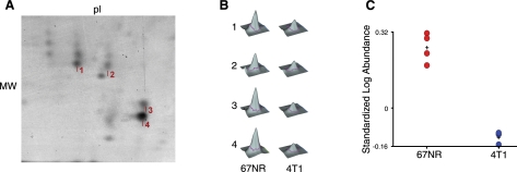
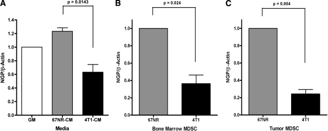
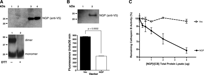
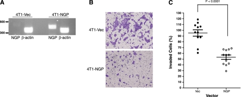
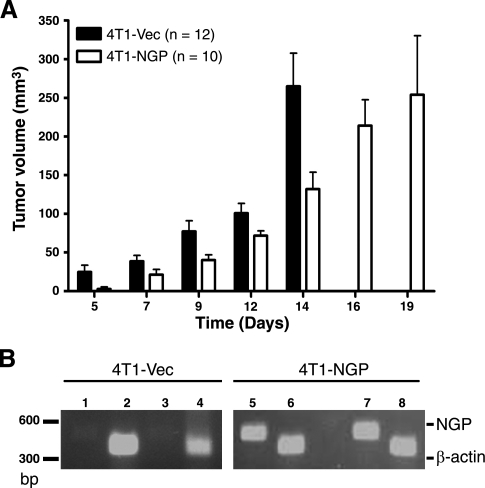
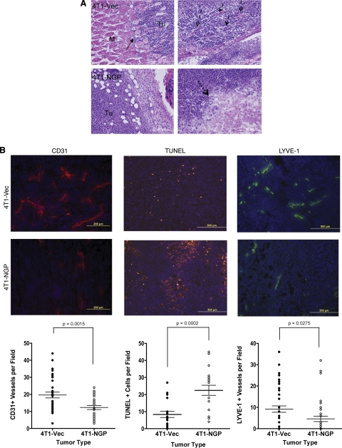
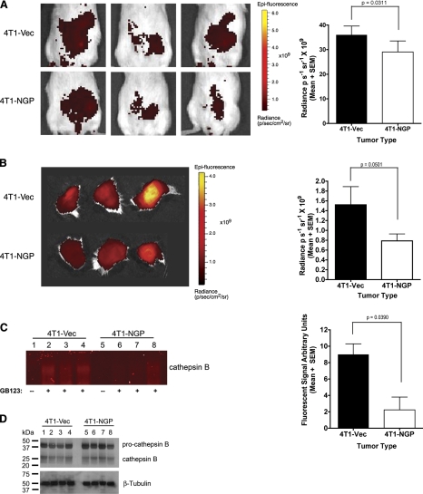
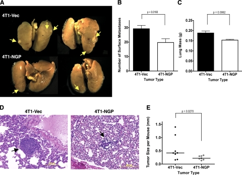
Myeloid-derived suppressor cells (MDSCs) are significantly increased in cancer patients and tumor bearing-animals. MDSCs infiltrate into tumors and promote tumor invasion and metastasis. To identify the mediator responsible for the prometastatic property of MDSCs, we used proteomics. We found neutrophilic granule protein (NGP) was decreased >2-fold in MDSCs from metastatic 4T1 tumor-bearing mice compared to nonmetastatic 67NR controls. NGP mRNA levels were decreased in bone marrow and in tumor-infiltrating MDSCs by 45 and 66%, respectively, in 4T1 tumor-bearing mice compared to 67NR controls. Interestingly, 4T1-conditioned medium reduced myeloid cell NGP expression by ∼ 40%, suggesting that a secreted factor mediates gene reduction. Sequence analysis shows a putative cystatin domain in NGP, and biochemical analysis confirms NGP a novel cathepsin inhibitor. It inhibited cathepsin B activity by nearly 40% in vitro. NGP expression in 4T1 tumor cells suppressed cell invasion, delayed primary tumor growth, and greatly reduced lung metastasis in vivo. A 2.8-fold reduction of cathepsin activity was found in tumors expressing NGP compared to controls. NGP significantly reduced tumor angiogenesis to 12.6 from 19.6 and lymphangiogenesis to 4.6 from 9.1 vessels/field. Necrosis was detectable only in NGP-expressing tumors, and the number of apoptotic cells increased to 22.4 from 8.3 in controls. Taken together, this study identifies a negative regulator of tumor metastasis in MDSCs, NGP, which is down-regulated in metastatic conditions. The finding suggests that malignant tumors promote invasion/metastasis not only through up-regulation of proteases but also down-regulation of protease inhibitors.


Figures

Figure 1.
Figure 1.
DIGE of NGP in MDSC lysates. Purified Gr1+/CD11b+ MDSCs isolated from spleen of mice bearing 4T1 tumors and 67NR tumors were analyzed by 2-dimensional gel electrophoresis. A) Representative image of a Sypro ruby-stained replicate gel for the migratory region of NGP. Protein spots 1–4 represent the NGP protein in the range of ∼20 kDa and pI ∼5. B) Peak intensity illustrations of peptide AIEAYNQGR from NGP isolated from 4 comigrating spots indicated in A. C) Graphical representation of the standardized log abundance of NGP protein spots 1–4 indicated inA. P = 0.000028 by Student's t test; P = 0.0014 with Benjamini-Hochberg false discovery rate correction. MDSC cell lysates were pooled from 6–7 mice/group and analyzed with 4 replicate gels.
Figure 2.
Figure 2.
Malignant tumors down-regulate NGP expression in myeloid cells and in MDSCs from bone marrow or tumor tissues. A) 32D cells were incubated with conditioned medium collected from 67NR or 4T1 cells or with growth medium (GM) for 24 h. NGP transcript levels were analyzed by real-time PCR. Data represent average ± sem ΔΔCT values compared to untreated cells. Each experiment was completed in triplicate and repeated twice. Results are significant by 1-tailed t test as indicated by P values. B, C) Gr1/CD11b cells were isolated from bone marrow (B) and tumor tissues (C) of mice bearing similar size 67NR tumors or 4T1 tumors by magnetic sorting (n=6 mice/group). Data represent average ± sem ΔΔCT values compared to MDSCs derived from 67NR tumor-bearing animals. RNA was isolated for real-time PCR to determine NGP expression. Experiments were repeated twice. All data were significant by 2-tailed paired t test, P < 0.05.
Figure 3.
Figure 3.
NGP inhibits cathepsin B activity in vitro. A) NIH-3T3 cells were transfected with empty (lanes 1 and 2) or NGP (lanes 3 and 4) expression vectors for 48 h, respectively. Cell lysate (lanes 1 and 3) and culture medium (lanes 2 and 4) were analyzed for NGP expression by Western blot using V5 antibody (top panel). NGP vector was transfected into HEK 293T for 48 h. Culture medium was analyzed for NGP expression by Western blot without (lane 1) or with (lane 2) 200 μM DTT (bottom panel). B) HEK 293T cells were transfected with Vec control (lane 1) or NGP vectors (lane 2) for 48 h. NGP expression was detected in culture medium (top panel). Endogenous cathepsin B activity was measured by fluorogenic substrate RR-AMC. P = 0.0002; 2-tailed t test (bottom panel). C) HEK 293T cells were transfected with either NGP or cathepsin B expression vectors, respectively, for 48 h. Cell lysates were mixed at the ratios indicated. Vec was used as control. Hydrolysis of cathepsin B fluorogenic substrate RR-AMC was measured. Percentage of remaining activity was calculated against the sample without NGP. P < 0.0001; 2-way ANOVA. Data are means ± se of duplicate experiments measured in triplicate.
Figure 4.
Figure 4.
NGP inhibits tumor cell invasion in vitro. A) RT-PCR of 4T1-Vec and 4T1-NGP stable cells. NGP (NGP-V5) is detectable at 460 bp. β-Actin at 380 bp was used as a loading control. B) Cells (8×10⁴) were plated onto Matrigel-coated Transwell inserts and incubated for 16 h. Cells that invaded through Matrigel-coated Transwell inserts were stained with crystal violet and photographed (×400). Images are representative. C) Quantification of invading cells. Data represent mean ± se percentage of invasion of 3 independent experiments performed in quadruplicate Transwell inserts. P < 0.0001; 2-tailed t test.
Figure 5.
Figure 5.
NGP inhibits tumor growth in vivo. A) 4T1-NGP or 4T1-Vec cells (5×10⁵) were injected into the fourth mammary fat pad of 6–7 wk old female Balb/c mice. Tumor volume was determined based on caliper measurements. Data represent mean ± se volume. P < 0.0001; 2-way ANOVA. B) Confirmation of NGP expression in primary tumors by RT-PCR. Lanes 1–4: 4T1-Vec; lanes 5–8: 4T1-NGP. Lanes 1, 3, 5, 7: NGP; lanes 2, 4, 6, 8: β-actin. NGP (NGP-V5) is indicated at 460 bp and β-actin at 380 bp.
Figure 6.
Figure 6.
NGP inhibits tumor angiogenesis, lymphangiogenesis, and invasion. A) Size-matched tumors were stained with H&E. Solid arrows indicate invasion of tumor cells into adjacent muscle tissue (top left panel). M, muscle; Tu, tumor. Dashed arrows indicate collagen fibers and desmoplasia the 4T1-Vec group (top right panel). Dotted arrows indicate a large necrotic lesion in 4T1-NGP tumor (bottom right panel). Images are representative. M, muscle; Tu, tumor. B) Representative images and quantitation of CD31 immunofluorescence, TUNEL fluorescent assay, or LYVE-1 immunofluorescence of size-matched tumors from 5–6 mice/group. Numbers of fields per section analyzed are indicated. Left panels: CD31+ vessels were counted in 3 fields/section. P = 0.0015; 2-tailed t test. Middle panels: 3 fields/section were counted for TUNEL assay to quantitate apoptotic cells. P = 0.0002; 2-tailed t test. Right panels: 5 fields/section were counted for LYVE-1+ vessels to quantify lymphatic vessels. P = 0.0275; 2-tailed, unpaired t test.
Figure 7.
Figure 7.
NGP inhibits cathepsin activity in vivo. A) Mice bearing size-matched 4T1 tumors were injected intravenously with probe GB123 at 18 h prior to in vivo imaging. Left panels: representative images. Right panel: photon intensity quantitation. P = 0.0311; t test. B) Left panel: whole tumors were removed surgically and imaged for cathepsin activity by GB123 probe intensity. Right panel: quantification of data. P = 0.05; t test. C) Left panel: equal amounts of total protein from tumor lysates were separated by PAGE; probe fluorescence at cathepsin B molecular weight was visualized on a flatbed scanner. Lane 1–4: 4T1-Vec, lane 1 without probe and lanes 2–4 with probe; lanes 5–8: 4T1-NGP, lane 5 without probe and lanes 6–8 with probe. Right panel: intensities of the labeled bands were quantified and plotted as means ± se. P = 0.039; t test. D) Protein samples identical to those in C were analyzed by Western blot for cathepsin B protein. β-Tubulin was used as a loading control. Each analysis utilized size-matched tumors from 3 mice/group.
Figure 8.
Figure 8.
NGP expression inhibits pulmonary metastasis of mammary tumors. A) Representative images of lung metastases after fixation in Bouin's fixative. Arrows indicate visible lung surface metastases. B) Number of visible surface metastases of lung tissues. P = 0.0160; 2-tailed t test. C) Measured weight of freshly harvested lungs. P = 0.0062; 2-tailed t test. D) Representative images of H&E-stained sections of lung tissues. E) Quantification of metastatic tumor diameter from H&E-stained sections. P = 0.0242; 2-tailed Welch's t test.

References

    1. Gabrilovich D. I., Nagaraj S. (2009) Myeloid-derived suppressor cells as regulators of the immune system. Nat. Rev. Immunol. 9, 162–174 - PMC - PubMed
    1. Almand B., Clark J. I., Nikitina E., van Beynen J., English N. R., Knight S. C., Carbone D. P., Gabrilovich D. I. (2001) Increased production of immature myeloid cells in cancer patients: a mechanism of immunosuppression in cancer J. Immunol. 166, 678–689 - PubMed
    1. Yang L., DeBusk L. M., Fukuda K., Fingleton B., Green-Jarvis B., Shyr Y., Matrisian L. M., Carbone D. P., Lin P. C. (2004) Expansion of myeloid immune suppressor Gr+CD11b+ cells in tumor-bearing host directly promotes tumor angiogenesis. Cancer Cell 6, 409–421 - PubMed
    1. Diaz-Montero C. M., Salem M. L., Nishimura M. I., Garrett-Mayer E., Cole D. J., Montero A. J. (2009) Increased circulating myeloid-derived suppressor cells correlate with clinical cancer stage, metastatic tumor burden, and doxorubicin-cyclophosphamide chemotherapy. Cancer Immunol. Immunother. 58, 49–59 - PMC - PubMed
    1. Shojaei F., Wu X., Malik A. K., Zhong C., Baldwin M. E., Schanz S., Fuh G., Gerber H.-P., Ferrara N. (2007) Tumor refractoriness to anti-VEGF treatment is mediated by CD11b+Gr1+ myeloid cells. Nat. Biotechnol. 25, 911–920 - PubMed

Publication types

MeSH terms