. 2011 May 10;108(19):7944-9.
doi: 10.1073/pnas.1100552108. Epub 2011 Apr 25.

IFN-lambda determines the intestinal epithelial antiviral host defense

Affiliations

IFN-lambda determines the intestinal epithelial antiviral host defense

Johanna Pott et al. Proc Natl Acad Sci U S A. .

Abstract

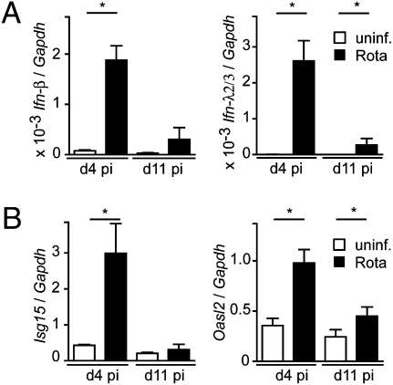
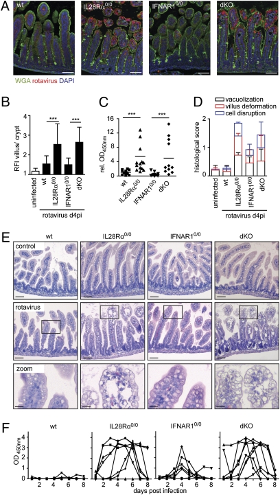
Type I and type III IFNs bind to different cell-surface receptors but induce identical signal transduction pathways, leading to the expression of antiviral host effector molecules. Despite the fact that type III IFN (IFN-λ) has been shown to predominantly act on mucosal organs, in vivo infection studies have failed to attribute a specific, nonredundant function. Instead, a predominant role of type I IFN was observed, which was explained by the ubiquitous expression of the type I IFN receptor. Here we comparatively analyzed the role of functional IFN-λ and type I IFN receptor signaling in the innate immune response to intestinal rotavirus infection in vivo, and determined viral replication and antiviral gene expression on the cellular level. We observed that both suckling and adult mice lacking functional receptors for IFN-λ were impaired in the control of oral rotavirus infection, whereas animals lacking functional receptors for type I IFN were similar to wild-type mice. Using Mx1 protein accumulation as marker for IFN responsiveness of individual cells, we demonstrate that intestinal epithelial cells, which are the prime target cells of rotavirus, strongly responded to IFN-λ but only marginally to type I IFN in vivo. Systemic treatment of suckling mice with IFN-λ repressed rotavirus replication in the gut, whereas treatment with type I IFN was not effective. These results are unique in identifying a critical role of IFN-λ in the epithelial antiviral host defense.


Conflict of interest statement

The authors declare no conflict of interest.

Figures

Fig. 1.
Fig. 1.
IECs express type I and III IFN genes after rotavirus challenge. Suckling C57BL/6 mice were orally infected with murine rotavirus strain EDIM (5 μL of 1:100 diluted virus stock). IECs were isolated at day 4 (n = 5) and day 11 (n = 5) postinfection (pi, black bars) or from uninfected control mice (open bars, n = 4) and analyzed by quantitative RT-PCR for expression of (A) Ifn-β, Ifn-λ2/3, and (B) Isg15 and Oasl2 genes. Results are representative for at least two independent experiments.
Fig. 2.
Fig. 2.
Enhanced rotavirus replication and IEC damage in mice lacking functional IFN-λ receptors. (A–E) Suckling wild-type (n = 12), IL28Rα0/0 (n = 14), IFNAR10/0 (n = 14), and IFNAR10/0IL28Rα0/0 (dKO, n = 12) mice were orally infected with murine rotavirus strain EDIM (5 μL of 1:100 diluted stock). Animals were killed 4 d later. (A) Immunostaining for rotavirus antigen (red) in thin sections of paraffin-embedded intestinal tissue. Counterstaining was performed with wheat germ agglutinin (WGA, green) and DAPI (blue). (Scale bars, 50 μm.) (B) Epithelial rotavirus antigen staining intensity was measured to obtain the relative fluorescence intensity (RFI) of villus/crypt epithelium. (C) Viral antigen levels in colon homogenates were determined by ELISA. Combined results of three independent experiments are shown. To facilitate comparison, OD450nm readings were normalized to the mean values obtained from wild-type mice of each experiment. (D) Histological score of virus-induced small intestinal tissue alterations and (E) H&E stainings of rotavirus-infected tissue. [Scale bars, 50 μm (Top and Middle), 10 μm (Bottom).] (F) Virus shedding in feces of adult wild-type, IL28Rα0/0, IFNAR10/0, and IFNAR10/0IL28Rα0/0 (dKO) mice (n = 8 for each group) after oral infection with murine rotavirus strain EDIM. Each line represents the OD450nm values of one individual mouse during the observed time period.
Fig. 3.
Fig. 3.
IECs of infected mice mainly respond to virus-induced IFN-λ. Intestinal tissue from rotavirus-infected suckling wild-type, IL28Rα0/0, IFNAR10/0, and IFNAR10/0IL28Rα0/0 (dKO) mice was harvested at day 4 postinfection. Paraffin-embedded samples were subjected to simultaneous staining for Mx1 (green), rotavirus antigen (white), and E-cadherin (red). Counterstaining was performed with DAPI (blue). (Right) Zoom column represents larger magnifications of the adjacent boxed areas. (Scale bars, 50 μm in first three columns; 20 μm in the zoom column.)
Fig. 4.
Fig. 4.
IFN-λ induces an epithelial cell-specific response. (A and B) The spatial response to in vivo electroporation of plasmids encoding mouse IFN-λ or mouse IFN-α was monitored by immunofluorescence staining for Mx1 in duodenal tissue sections of (A) IFNAR10/0 or IL28Rα0/0 mice expressing IFN-α, and (B) in IL28Rα0/0 and IFNAR10/0 mice expressing IFN-λ. (Scale bar, 50 μm.) (C–E) Stat1 tyrosine phosphorylation was detected by immunoblotting after stimulation with IFN (human IFN-αB/D, 2,000 U/mL; mouse IFN-β, 500 U/mL; or mouse IFN-λ, 20 ng/mL). (C) IECs isolated from adult C57BL/6 wild-type mice were stimulated ex vivo for 1 h with the indicated IFN. Unstimulated IECs served as negative control. The images show triplicates for each condition. (D) Mouse macrophage-like RAW 264.7 cells were stimulated for the indicated time. (E) Rat intestinal epithelial IEC-6 cells were grown to confluency on transwell filters and left untreated or stimulated either apically or basolaterally with the indicated IFN. Actin staining was included to demonstrate equal protein loading. The images are representative of three independent experiments.
Fig. 5.
Fig. 5.
Administration of IFN-λ mediates rotavirus protection. Suckling wild-type mice (10 animals per group) were given subcutaneous injections of mouse IFN-λ2 (1 μg per injection), human hybrid IFN-αB/D (1 μg per injection), or buffer only (mock) 8 h before oral infection with (A) high dose (5 μL of 1:103 diluted virus stock) or (B) low dose (5 μL of 1:104 diluted virus stock) of murine rotavirus strain EDIM. The IFN treatment was repeated on days 1 and 2 postinfection. OD450nm values representing the viral antigen levels in colon homogenates on day 3 postinfection are shown.

Similar articles

Cited by

References

    1. Perry AK, Chen G, Zheng D, Tang H, Cheng G. The host type I interferon response to viral and bacterial infections. Cell Res. 2005;15:407–422. - PubMed
    1. Kotenko SV, et al. IFN-lambdas mediate antiviral protection through a distinct class II cytokine receptor complex. Nat Immunol. 2003;4:69–77. - PubMed
    1. Sheppard P, et al. IL-28, IL-29 and their class II cytokine receptor IL-28R. Nat Immunol. 2003;4:63–68. - PubMed
    1. Gad HH, et al. Interferon-lambda is functionally an interferon but structurally related to the interleukin-10 family. J Biol Chem. 2009;284:20869–20875. - PMC - PubMed
    1. Onoguchi K, et al. Viral infections activate types I and III interferon genes through a common mechanism. J Biol Chem. 2007;282:7576–7581. - PubMed

Publication types

MeSH terms