

Volumetric abnormalities predating the onset of schizophrenia and affective psychoses: an MRI study in subjects at ultrahigh risk of psychosis
- PMID: 21518921
- PMCID: PMC3446227
- DOI: 10.1093/schbul/sbr035
Volumetric abnormalities predating the onset of schizophrenia and affective psychoses: an MRI study in subjects at ultrahigh risk of psychosis
Abstract
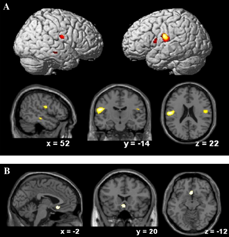
It remains unclear whether brain structural abnormalities observed before the onset of psychosis are specific to schizophrenia or are common to all psychotic disorders. This study aimed to measure regional gray matter volume prior to the onset of schizophreniform and of affective psychoses. We investigated 102 subjects at ultrahigh risk (UHR) of developing psychosis recruited from the Personal Assessment and Crisis Evaluation Clinic in Melbourne, Australia. Twenty-eight of these subjects developed psychosis subsequent to scanning: 19 schizophrenia, 7 affective psychoses, and 2 other psychoses. We examined regional gray matter volume using 1.5 mm thick, coronal, 1.5 Tesla magnetic resonance imaging and voxel-based morphometry methods of image analysis. Subjects were scanned at presentation and were followed up clinically for a minimum of 12 months, to detect later transition to psychosis. We found that both groups of subjects who subsequently developed psychosis (schizophrenia and affective psychosis) showed reductions in the frontal cortex relative to UHR subjects who did not develop psychosis. The subgroup that subsequently developed schizophrenia also showed smaller volumes in the parietal cortex and, at trend level, in the temporal cortex, whereas those who developed an affective psychosis had significantly smaller subgenual cingulate volumes. These preliminary findings suggest that volumetric abnormalities in UHR individuals developing schizophrenia vs affective psychoses comprise a combination of features that predate both disorders and others that may be specific to the nature of the subsequent disorder.
Figures
References
-
- Haldane M, Frangou S. New insights help define the pathophysiology of bipolar affective disorder: neuroimaging and neuropathology findings. Prog Neuropsychopharmacol Biol Psychiatry. 2004;28:943–960. - PubMed
-
- Koo MS, Levitt JJ, Salisbury DF, Nakamura M, Shenton ME, McCarley RW. A cross-sectional and longitudinal magnetic resonance imaging study of cingulate gyrus gray matter volume abnormalities in first-episode schizophrenia and first-episode affective psychosis. Arch Gen Psychiatry. 2008;65:746–760. - PMC - PubMed
-
- Tang Y, Wang F, Xie G, et al. Reduced ventral anterior cingulate and amygdala volumes in medication-naive females with major depressive disorder: a voxel-based morphometric magnetic resonance imaging study. Psychiatry Res. 2007;156:83–86. - PubMed
-
- Baare WF, van Oel CJ, Hulshoff Pol HE, et al. Volumes of brain structures in twins discordant for schizophrenia. Arch Gen Psychiatry. 2001;58:33–40. - PubMed
Publication types
MeSH terms
Grants and funding
LinkOut - more resources
Full Text Sources
Medical
