. 2011 Jul;31(13):2729-41.
doi: 10.1128/MCB.05165-11. Epub 2011 Apr 25.

A barrier-only boundary element delimits the formation of facultative heterochromatin in Drosophila melanogaster and vertebrates

Affiliations

A barrier-only boundary element delimits the formation of facultative heterochromatin in Drosophila melanogaster and vertebrates

Nianwei Lin et al. Mol Cell Biol. 2011 Jul.

Abstract

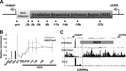
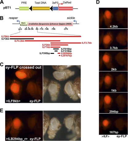
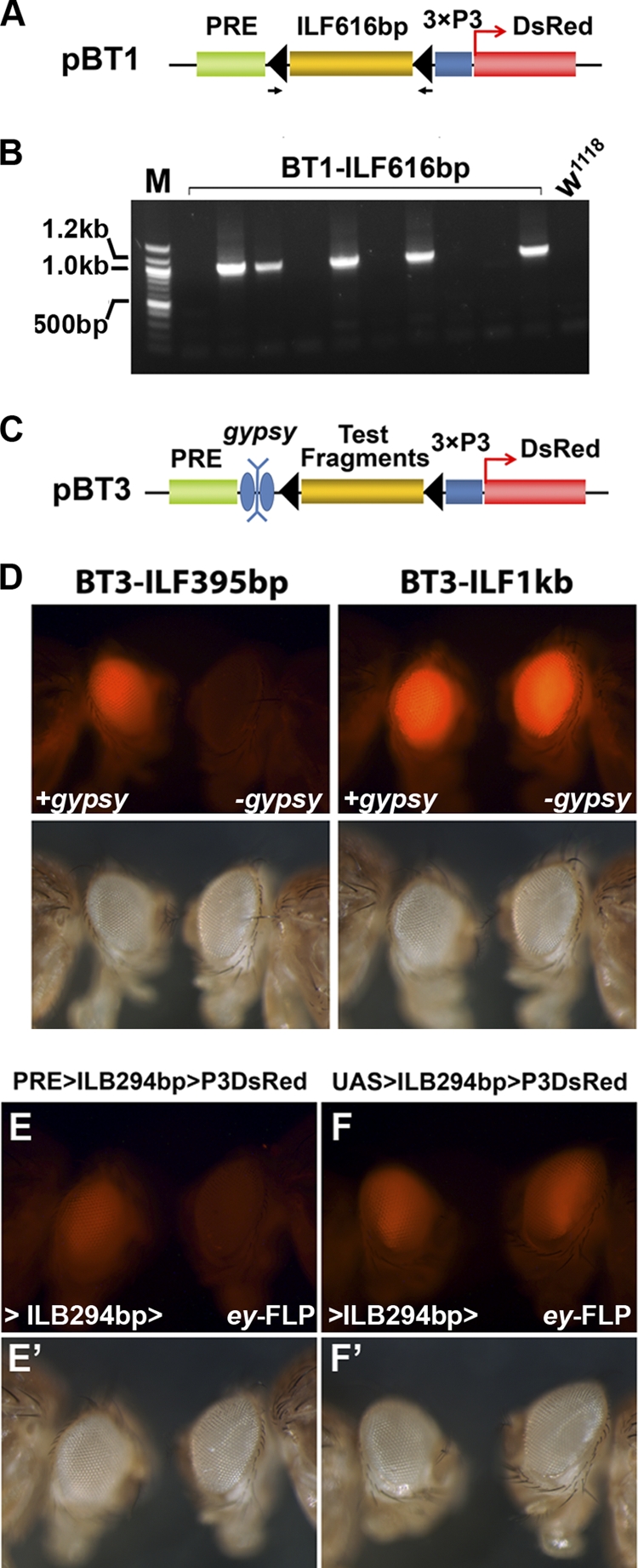
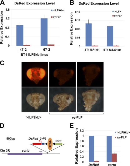
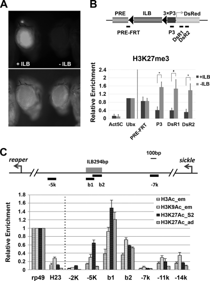
Formation of facultative heterochromatin at specific genomic loci is fundamentally important in defining cellular properties such as differentiation potential and responsiveness to developmental, physiological, and environmental stimuli. By the nature of their formation, heterochromatin and repressive histone marks propagate until the chain reaction is broken. While certain active promoters can block propagation of heterochromatin, there are also specialized DNA elements, referred to as chromatin barriers, that serve to demarcate the boundary of facultative heterochromatin formation. In this study, we identified a chromatin barrier that specifically limits the formation of repressive chromatin to a distal enhancer region so that repressive histone modifications cannot reach the promoter and promoter-proximal enhancer regions of reaper. Unlike all of the known boundary elements identified for Drosophila melanogaster, this IRER (irradiation-responsive enhancer region) left barrier (ILB) does not exhibit enhancer-blocking activity. Not only has the ILB been conserved in different Drosophila species, it can also function as an effective chromatin barrier in vertebrate cells. This suggests that the mechanism by which it functions to spatially restrict the formation of repressive chromatin marked by trimethylated H3K27 has also been conserved widely during evolution.


Figures

Fig. 1.
Fig. 1.
Formation of facultative heterochromatin is restricted to the IRER, without reaching the reaper promoter and proximal enhancer regions. (A) Schematic diagram of the intergenic region between reaper and sickle. The IRER is required to mediate irradiation-induced expression of the proapoptotic genes reaper, hid, and sickle (54). Accessibility of the IRER is controlled by a PcG protein-dependent mechanism, which forms a nonpermissive structure in irradiation-resistant cells in post-stage-12 embryos. The decrease of DNA accessibility, accompanied by enrichment of repressive histone marks and binding of PcG proteins, was specifically limited to the IRER without affecting the reaper promoter and proximal enhancer region (54). (B) ChIP assays performed with wild-type adult fly tissues. Pericentromeric heterochromatin locus H23 (H23) and the Ubx promoter region (Ubx) were used as positive controls for repressive histone marks H3K9me3 and H3K27me3, respectively. The coding region of the housekeeping gene Act5C was used as a background control. Enrichment of the repressive histone marks in the IRER was normalized against the respective positive controls and is presented as means ± standard deviations (SD). The high-level enrichment of both H3K27me3 and H3K9me3 in the central part of the IRER dropped significantly at the left boundary of the IRER, about −2 kb to −5 kb relative to the reaper TSS. (C) Distribution of histone marks H3K4me3 and H3K27me3 and binding of Pol II in Drosophila S2 cells revealed by ChIP-Seq. The relative locations of reaper and sickle are indicated by arrows. The dotted vertical line denotes the region that might possess putative chromatin barrier activity.
Fig. 2.
Fig. 2.
Verification of barrier activity and narrowing down the IRER left barrier. (A) Barrier tester construct pBT1. Eye-specific 3×P3-DsRed served as the reporter/marker. The PRE from the Ubx promoter was placed upstream of 3×P3-DsRed to initiate the formation of facultative heterochromatin. The tested fragments (ILFs) were cloned between the reporter gene and the PRE and were flanked by two FRT sequences. The transgenic flies were generated by either P-mediated insertion or ΦC31-mediated integration. (B) A series of fragments within the IRER left boundary region were tested with the pBT1 vector for barrier activity. The fragments with and without barrier activity are shown as red and black bars, respectively. The essential barrier region was narrowed down to the ILB167bp region. (C) Example of verification of barrier activity. The left and right panels show the same group of flies under the RFP fluorescence channel and bright-field microscopy, respectively. Transgenic line 47-2, carrying one copy of pBT1-ILF9kb (>ILF9kb>; fly head on the left), had a strong eye-specific DsRed signal, which was diminished when the ILF9kb fragment was removed by crossing with an ey-FLP strain (ey-FLP; fly head on the right). The DsRed signal was fully restored when ey-FLP was crossed out (top fly head). (D) pBT1 constructs carrying subfragments of ILF9kb were integrated into the same attP docking site on the 2nd chromosome. Barrier activity was verified as mentioned above. The fly heads of the original transgenic strains are on the left, while those that also have ey-FLP are on the right side of each panel. This series of tests indicated that the ILB167 fragment possessed full barrier activity compared to longer fragments. (E) The barrier activity was not affected when the ILB294bp fragment was inserted into pBT1 in the reverse direction, indicating that ILB is orientation independent.
Fig. 3.
Fig. 3.
Verification of tested fragments. (A and B) PCR verification of the transformation events demonstrated with BT1-ILF616bp, from which no DsRed-positive flies were recovered. Randomly selected progenies from each individual vial were collected for genomic DNA extraction, and PCR analysis was performed with a pair of primers flanking the two FRT sequences. The genomic DNAs from 5 of 10 tested vials (each established with 2 or 3 injected adults) showed PCR products of around 1 kb, indicating an ∼50% transformation rate. This evidence indicates that the failure to recover BT1-ILF616bp (and other negative fragments) was not due to potential problems associated with transformation but rather was due to the silencing of the reporter gene by the PRE, i.e., the lack of barrier activity of the tested fragment. (C) Some of the negative fragments were further verified with the reporter construct pBT3, which contains a gypsy element flanked by two loxP sequences between the PRE and the test DNA fragment. Transgenic flies were generated with the ΦC31 line 9752. Germ line excision of the gypsy element was performed by crossing the transgenic flies to a strain providing the source of Cre recombinase (y w; Sco/CyO Crew1). ILF395bp was negative, while ILF1kb tested positive in the original BT1-mediated assay. (D) Both BT3-ILF395bp and BT3-ILF1kb transgenic files had similar levels of DsRed in the presence of the gypsy insulator (+gypsy). However, the level of DsRed in the BT3-ILF395bp line diminished after the excision of gypsy (left panel, −gypsy), indicating that ILF395bp does not have barrier activity. In contrast, the excision of the gypsy insulator from BT3-ILF1kb did not lead to any detectable decrease of the DsRed signal (right panel, −gypsy), indicating that ILF1kb is sufficient to block heterochromatin formation initiated by the PRE. To verify that ILB294bp does not have eye-specific enhancer activity, transgenic lines carrying BT1-ILB294 (PRE>ILB294bp>P3DsRed) (E and E′) or IT1-ILB294 (UAS>ILB294bp>P3DsRed) (F and F′) were crossed to flies carrying ey-FLP. The BT1-ILB294 transformant line showed a significant reduction of DsRed signal after the somatic excision mediated by ey-FLP. In contrast, flies carrying IT1-ILB294bp had little change after being crossed with ey-FLP. This indicates that there is no eye-specific enhancer activity associated with the 294-bp fragment.
Fig. 4.
Fig. 4.
ILB prevents transcriptional silencing mediated by PRE. (A and B) The mRNA level of the 3×P3-DsRed reporter gene, detected by Q-PCR, was significantly reduced after the excision of the ILB-containing fragments by ey-FLP. BT1-ILF9kb transgenic lines 47-2 and 67-2 were generated by P insertions (A), while the BT1-ILF1kb and BT1-ILB294bp lines were generated by ΦC31-mediated integration (B). (C) When crossed to ey-FLP, in addition to the decreased DsRed signal, the BT1-ILF9kb transgenic line 67-2 showed an eye ablation phenotype similar to that of a corto mutant. (D) Inverse PCR identified that the BT1-ILF9kb transgene in line 67-2 was inserted about 400 bp upstream of the corto gene by P insertion. (E) For line 67-2, the level of corto expression was significantly reduced, to less than 50% of the original level, after the excision of ILF9kb by ey-FLP.
Fig. 5.
Fig. 5.
ILB blocks the propagation of repressive histone marks initiated by the PRE. (A) hs-FLP was used to remove the ILB-containing fragment through germ line recombination. No DsRed signal was detectable in the resulting PRE>3×P3-DsRed flies (−ILB). (B) Enrichment of H3K27me3 in and around the reporter gene before (+ILB) and after (−ILB) the removal of ILB via germ line recombination. Targeted loci for primer pairs for the ChIP assays are indicated by black bars below the schematic map of the transgene. Removal of ILB led to significant enrichment of H3K27me3 in the reporter gene loci P3, DsR1, and DsR2 (*, P < 0.05; #, P = 0.06847). Note that the level of H3K27me3 remained about the same for the PRE-FRT locus, which is not shielded by ILB. (C) Higher levels of histone H3 acetylation at the barrier site. The ILB294bp region (b1 and b2, approximately 6 kb from the reaper TSS) has a significantly higher level of H3 acetylation than the surrounding region. Specifically, both H3K9 and H3K27 are hyperacetylated in the ILB294 region. ChIP assays were performed with late-stage embryos (H3Ac and H3K9Ac), S2 cells (H3K27Ac), and adult flies (H3K27Ac). Data were normalized against the recovery rate for the rp49 locus before statistical analysis.
Fig. 6.
Fig. 6.
The IRER left barrier does not contain enhancer-blocking activity. (A) The reporter construct pIT1 was designed to test enhancer-blocking activity. DNA fragments flanked by FRT were inserted between a UAS sequence and the 3×P3-DsRed reporter. Transgenic flies carrying pIT1 can be crossed to an engrailed (en)-GAL4 UAS-GFP strain. (B) If the DNA fragment has enhancer-blocking activity, such as the gypsy insulator, DsRed cannot be expressed in the engrailed pattern. (B′) GFP channel showing the same larva representing expression of en-GAL4. (C and C′) When the gypsy insulator was removed by FLP, DsRed was expressed in the same engrailed pattern as GFP. (D) With this testing scheme, the ILB294bp barrier element, which had complete barrier activity, did not display any detectable enhancer-blocking activity. (E to J) Potential enhancer-blocking activity of ILB was also assayed with the pCfhL reporter system. The expression of lacZ was placed under the control of ftz UPS and NE enhancers for expression in early and later embryogenesis, respectively. While the 1.2-kb Fab-7 insulator completely blocked the enhancer function of UPS and NE (E and F), the 1.27-kb fragment encompassing the essential ILB294bp barrier sequences did not block the enhancer-promoter interaction for either UPS or NE (G to J). lacZ expression was detected by X-Gal (5-bromo-4-chloro-3-indolyl-β-d-galactopyranoside) staining in germ band-extended (stage 9/10) embryos (E, G, and I) or germ band-retracted (stage 13) embryos (F, H, and J). Five independent pCfhl-ILB transgenic lines were tested, and none blocked UPS or NE enhancer function.
Fig. 7.
Fig. 7.
Cut is required for ILB activity. (A) The ILB167bp fragment contains a putative CDP/Cut binding site (large capital letters). The logo representation of the V$CDP_02 matrix is aligned on the bottom and is composed of a palindromic ATCGAT motif (highlighted in red in the corresponding putative binding site) overlapping with the homeodomain binding motif ATTA (italic sequence). (B) Cut protein was highly enriched in the 300-bp region encompassing ILB294bp (b1 and b2), as shown by ChIP analysis of both S2 cells and adult flies. (C) BT1-ILB294bp homozygous females were crossed to either w1118 males or ctC145 males, and the DsRed levels of their female progeny (aged for 2 days) are shown. Significantly decreased levels of DsRed were found in animals heterozygous for ctC145 compared to wild-type (wt) animals. (D) The Cut/CDP binding site in ILB is required for the barrier activity of ILB. When the binding site was mutated (in red), ILB_1k no longer had barrier activity following the removal of the gypsy insulator (for negative and positive examples, refer to Fig. 3). For both panels C and D, the left and right panels are pictures of the same flies taken with the DsRed filter set and no filter (regular light), respectively.
Fig. 8.
Fig. 8.
ILB is evolutionally conserved. (A) The 2-kb D. pseudoobscura genomic sequence encompassing the ILB294bp orthologous region (pseILB) displayed a similar level of barrier activity in D. melanogaster to that of native ILB. The transgenic flies were generated by ΦC31-mediated integration. (B) The 294-bp ILB protected the reporter from silencing in chicken erythroid 6C2 cells. The designs of the reporters used for generating stable transfection lines are shown on the left. Following hygromycin withdrawal, the reporter without barrier protection was silenced in most cells, as indicated by FACS analyses of IL-2R expression (top). However, when the reporter was flanked on both sides by one copy of ILB294bp (bottom), the expression of the reporter was maintained. Data for representative clones are shown. (C) Diagram summarizing the function of ILB. The binding of Cut likely recruits a histone acetyltransferase such as dCBP, which catalyzes a euchromatic histone modification that is incompatible with heterochromatin formation. The potential interaction between Cut and dCBP remains to be verified. The presence of other DNA binding proteins in addition to Cut is suggested by the conservation of other DNA motifs/binding sites within ILB.

References

    1. Andres V., Chiara M. D., Mahdavi V. 1994. A new bipartite DNA-binding domain: cooperative interaction between the cut repeat and homeo domain of the cut homeo proteins. Genes Dev. 8:245–257 - PubMed
    1. Angrisano T., et al. 2011. Chromatin and DNA methylation dynamics during retinoic acid-induced RET gene transcriptional activation in neuroblastoma cells. Nucleic Acids Res. 39:1993–2006 - PMC - PubMed
    1. Bell A. C., West A. G., Felsenfeld G. 1999. The protein CTCF is required for the enhancer blocking activity of vertebrate insulators. Cell 98:387–396 - PubMed
    1. Bello B. C., Hirth F., Gould A. P. 2003. A pulse of the Drosophila Hox protein Abdominal-A schedules the end of neural proliferation via neuroblast apoptosis. Neuron 37:209–219 - PubMed
    1. Bi X., Broach J. R. 2001. Chromosomal boundaries in S. cerevisiae. Curr. Opin. Genet. Dev. 11:199–204 - PubMed

Publication types

MeSH terms

LinkOut - more resources