. 2011 May 10;123(18):1953-62.
doi: 10.1161/CIRCULATIONAHA.110.988642. Epub 2011 Apr 25.

G protein-coupled receptor kinase 2 activity impairs cardiac glucose uptake and promotes insulin resistance after myocardial ischemia

Affiliations

G protein-coupled receptor kinase 2 activity impairs cardiac glucose uptake and promotes insulin resistance after myocardial ischemia

Michele Ciccarelli et al. Circulation. .

Abstract

Background: Alterations in cardiac energy metabolism downstream of neurohormonal stimulation play a crucial role in the pathogenesis of heart failure. The chronic adrenergic stimulation that accompanies heart failure is a signaling abnormality that leads to the upregulation of G protein-coupled receptor kinase 2 (GRK2), which is pathological in the myocyte during disease progression in part owing to uncoupling of the β-adrenergic receptor system. In this study, we explored the possibility that enhanced GRK2 expression and activity, as seen during heart failure, can negatively affect cardiac metabolism as part of its pathogenic profile.

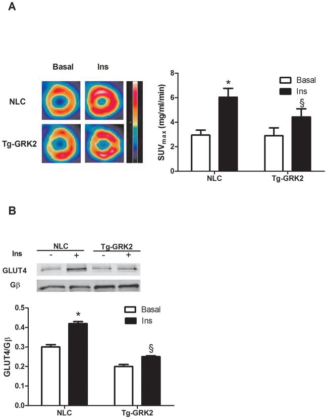
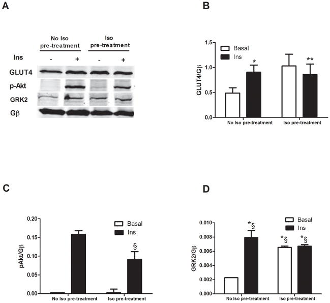
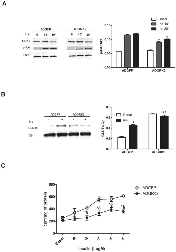
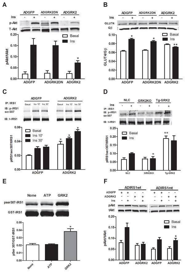
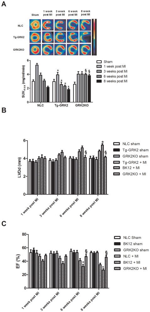
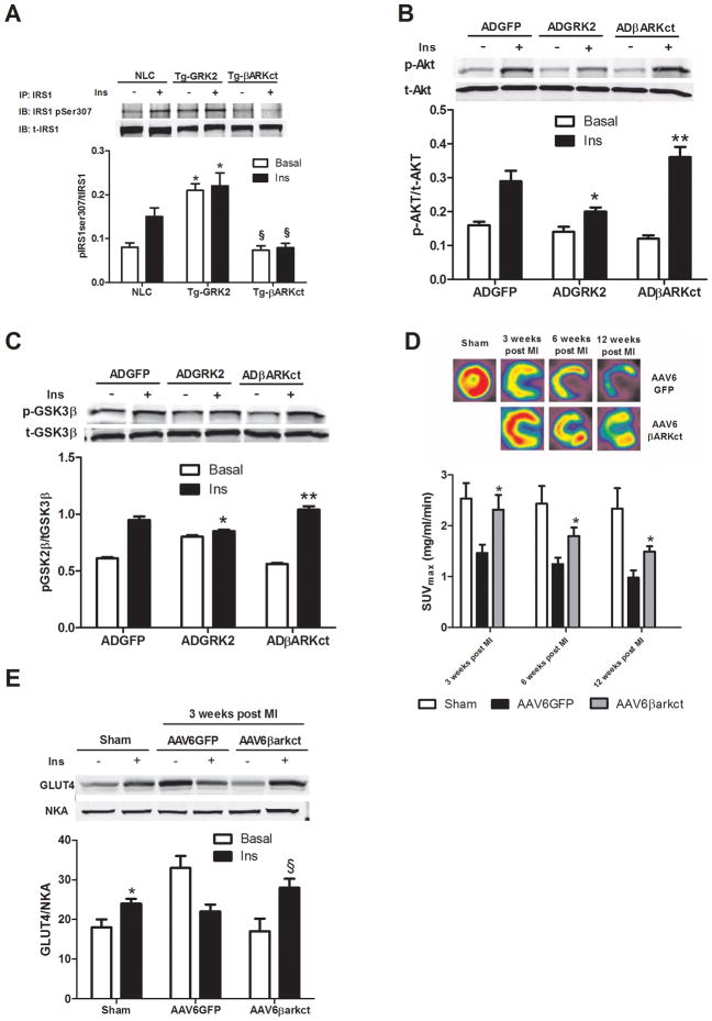
Methods and results: Positron emission tomography studies revealed in transgenic mice that cardiac-specific overexpression of GRK2 negatively affected cardiac metabolism by inhibiting glucose uptake and desensitization of insulin signaling, which increases after ischemic injury and precedes heart failure development. Mechanistically, GRK2 interacts with and directly phosphorylates insulin receptor substrate-1 in cardiomyocytes, causing insulin-dependent negative signaling feedback, including inhibition of membrane translocation of the glucose transporter GLUT4. This identifies insulin receptor substrate-1 as a novel nonreceptor target for GRK2 and represents a new pathological mechanism for this kinase in the failing heart. Importantly, inhibition of GRK2 activity prevents postischemic defects in myocardial insulin signaling and improves cardiac metabolism via normalized glucose uptake, which appears to participate in GRK2-targeted prevention of heart failure.

Conclusions: Our data provide novel insights into how GRK2 is pathological in the injured heart. Moreover, it appears to be a critical mechanistic link within neurohormonal crosstalk governing cardiac contractile signaling/function through β-adrenergic receptors and metabolism through the insulin receptor.


Figures

Figure 1
Figure 1. GRK2 levels influence in vivo cardiac glucose uptake
(A) Tg-GRK2 and NLC mice were studied by PET to evaluate cardiac glucose uptake after intraperitoneal injection of insulin (Ins, 0.075U/Kg, IP). Ins significantly increases glucose uptake however this response was attenuated in BK12 mice (*, P<0.05, Ins vs Basal, §, P<0.05, TG-GRK2 vs NLC, n=8 per group). (B) Plasma membrane GLUT4 protein levels from Tg-GRK2 and NLC hearts. Animals were injected with Ins as above and sacrificed 15 min later. GLUT4 levels on the plasma membrane were increased by Ins in NLC but attenuated in TG-GRK2. GLUT4 level was normalized to the membrane protein Gβ (*, P<0.05, Ins vs Basal, §, P<0.05, fold of increased translocation to the membrane, TG-GRK2 vs NLC, n=3 per group).
Figure 2
Figure 2. Interplay between βAR and insulin receptor signaling
ARVMs were stimulated with Ins (0.1 μM for 10 min) with or without pre-treatment with the βAR agonist isoproterenol (Iso, 10μM for 5 min). Membrane fractions were prepared and blotted for signaling proteins. (A) Representative western blot showing membrane GLUT4, pAkt and GRK2. Gβ blotting was used as loading control. (B–D) Bar Charts showing GLUT4 translocation (*, P<0.05 Ins vs. Basal, n=3; **, n.s, Iso+Ins vs Iso, n=3per group), Akt was activated in response to Ins but attenuated with Iso pretreatment (§ P<0.05, fold activation, Iso+Ins vs. Ins alone, n=3 per group) and GRK2 translocation induced by Ins with or without Iso pretreatment (*§, P<0.05 Ins, Iso and Iso+Ins vs Basal, n=3 per group).
Figure 3
Figure 3. GRK2 overexpression in ARVMs inhibits cellular effects of insulin
Adenoviral-infected ARVMs were treated with or without 0.1uM Ins for either 10 or 30min. (A) Representative Western blot from ARVMs whole lysate. Ins activation of Akt was attenuated by GRK2 overexpression. Total Akt was blotted as loading control (*, P<0.05 fold activation vs ADGFP, n=3 per group). (B) Western blot from plasma membranes of ARVM showing increased GLUT4 translocation in response to 0.1uM Ins for 10min with GFP overexpression but blunted with GRK overexpression (*, P<0.05 vs Basal; **, n.s. vs Basal, n=3), Gβ was used as loading control. (C) ARVMs were stimulated with Ins at different concentrations, ranging from 1 nM to 10 μM for 10 min in the presence of ADGFP or ADGRK2 infection. The rate of glucose uptake was determined by [3H] 2-DOG (*§, P < 0.01 vs. ADGFP, n=3 per group).
Figure 4
Figure 4. Mechanism of inhibitory effects of GRK2 on insulin signaling in myocytes
(A–B) ARVMs were infected with ADGFP, ADGRK2, or kinase-dead GRK2 (ADGRK2DN) and stimulated with Ins (0.1 μM for 10 min). (A) Representative immunoblot in whole cell lysates showing reduced pAkt level when GRK2 is overexpressed (*, fold activation, P<0.01 vs ADGFP and ADGRK2DN, n=3 per group). (B) Ins-stimulated GLUT4 membrane levels in ARVMs is attenuated with GRK2 overexpression (*, P<0.05 vs Basal; **, n.s. vs Basal, n=3) (C) IRS1 was immunoprecipitated from whole lysate of ARVMs infected with ADGFP or ADGRK2 and stimulated with Ins as above. Top panel, representative immunoblot for p-ser307 showing increased p-ser307 with ADGRK2 treatment. Lower panel is total IRS-1 (* P<0.05 vs ADGFP, n= 3). (D) Level of p-Ser307 of IRS1 was evaluated by immunoprecipitation of equal amount of IRS1 from isolated myocytes of NLC, Tg-GRK2 and cardiac GRK2 KO mice after stimulation with Ins (10−7 M, 10 min). Representative blots for GRK2, pSer307 IRS1 and total IRS1 are shown. Reduced and increased levels of pSer307IRS1 were respectively observed in GRK2KO and Tg-GRK2 versus NLC (*, P<0.01, fold of activation, GRK2KO vs NLC, n=3; **, P <0.01, Tg-GRK2 vs NLC, n=3 per group). (E) GRK2 phosphorylates IRS1 at Ser307. An in-vitro kinase assay was performed with purified GST-IRS1 alone (none) or in the presence of ATP or ATP plus purified GRK2. Representative blot from 3 independent assays (top panel) and densitometric analysis (lower panel) are shown. (*, P<0.05, fold activation vs ATP and none) (F) NRVMs were co-infected with either ADIRS1wt or ADIRS1 mutant Ser307Ala (mt) in addition to ADGFP or ADGRK2 and stimulated with Ins as above. Representative immunoblot showing p-Akt levels is provided (*, P<0.01, fold activation vs ADGRK2+ADIRS1wt, n=3 per group).
Figure 5
Figure 5. Cardiac glucose uptake in post-MI mice
(A) 18-FDG cardiac uptake in NLC Tg-GRK2 and cardiac-specific GRK2KO mice under Sham conditions and 1–8 weeks post-MI. Shown at top are representative micro-PET images where infarct is clearly visible and histogram below quantifies 18-FDG uptake in myocardium (*, P<0.05, TG-GRK2 vs NLC; §, P<0.05, GRK2KO vs NLC, n=12 per group). (B–C) LV diameter at diastole and Ejection fraction (EF) in NLC, Tg-GRK2 and GRK2KO mice determined by echocardiography serially after MI. (*, P<0.01, Tg-GRK2 vs NLC; §, P<0.01, GRK2KO vs NLC, n=12, per group).
Figure 6
Figure 6. Inhibition of GRK2 in myocytes and myocardium restores normal insulin signaling and prevents defective in vivo glucose uptake post-MI
(A) Level of Ins-stimulated pSer307 in IRS1 immunoprecipitated from adult mouse ventricular myocytes isolated from NLC, Tg-GRK2, or Tg-βARKct mice (*, P<0.01 vs NLC, n=3; §, P<0.01, Tg-βARKct vs NLC, n=3 per group). (B) ARVMs were infected with either ADGFP, ADGRK2 or ADβARKct. Representative blots for p-Akt and t-Akt are shown (*, P <0.01, fold increase in activation, ADGRK2 vs ADGFP, n=3;**, P <0.01, fold increase in activation, ADβARKct vs ADGRK2 and ADGFP, n=3 per group). (C) Representative western blot from ARVMs whole lysate infected as in (A) and stimulated with Ins showing pGSK3β and total-GSK3β (*, P <0.01, fold increase in activation, ADGRK2 vs ADGFP, n=3; **, P <0.01, fold increase in activation, ADβARKct vs ADGRK2, n=3). (D) Global rat myocardial in vivo glucose uptake evaluated by micro-PET in Sham and 1, 3, 6, and 12 weeks post-MI rats treated with AAV6-GFP or AAV6-βARKct (*, P<0.05, vs AAV6GFP, n=8 per group). (E) Rats 3 weeks post MI and Sham were sacrificed 15 min after an IP injection of insulin (0.075 U/Kg) and hearts removed. Plasma membranes were prepared to evaluate GLUT4 levels as shown in a representative blot (*, P<0.05 vs Basal; §, P< 0.05 vs Basal, n=3 per group).

References

    1. Braunwald E. The Denolin lecture. Congestive heart failure: a half century perspective. Eur Heart J. 2001;22:825–836. - PubMed
    1. Jessup M, Brozena S. Heart failure. N Engl J Med. 2003;348:2007–2018. - PubMed
    1. Neubauer S. The failing heart--an engine out of fuel. N Engl J Med. 2007;356:1140–1151. - PubMed
    1. Ingwall JS, Weiss RG. Is the failing heart energy starved? On using chemical energy to support cardiac function. Cir Res. 2004;95:135–145. - PubMed
    1. Ashrafian H, Frenneaux MP. Metabolic modulation in heart failure: the coming of age. Cardiovasc Drugs Ther. 2007;21:5–7. - PubMed

Publication types

MeSH terms