Comparative Study
. 2011 Jul;113(1):145-52.
doi: 10.1213/ANE.0b013e3182185fee. Epub 2011 Apr 25.

The potential dual effects of anesthetic isoflurane on hypoxia-induced caspase-3 activation and increases in β-site amyloid precursor protein-cleaving enzyme levels

Affiliations
Comparative Study

The potential dual effects of anesthetic isoflurane on hypoxia-induced caspase-3 activation and increases in β-site amyloid precursor protein-cleaving enzyme levels

Chuxiong Pan et al. Anesth Analg. 2011 Jul.

Abstract

Background: β-Amyloid protein (Aβ) accumulation, caspase activation, apoptosis, and hypoxia-induced neurotoxicity have been suggested to be involved in Alzheimer disease neuropathogenesis. Aβ is produced from amyloid precursor protein through proteolytic processing by the aspartyl protease β-site amyloid precursor protein-cleaving enzyme (BACE) and γ-secretase. Inhaled anesthetics have long been considered to protect against neurotoxicity. However, recent studies have suggested that the inhaled anesthetic isoflurane may promote neurotoxicity by inducing caspase activation and apoptosis, and by increasing levels of BACE and Aβ. We therefore sought to determine whether isoflurane can induce concentration-dependent dual effects on hypoxia-induced caspase-3 activation and increases in BACE levels: protection versus promotion.

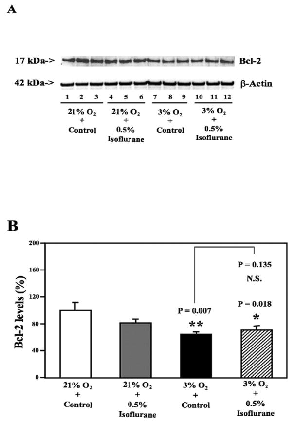
Methods: H4 human neuroglioma cells were treated with hypoxia (3% O(2)) alone, different concentrations of isoflurane (0.5% and 2%), and the combination of hypoxia and 0.5% or 2% isoflurane. The levels of caspase-3 cleavage (activation), BACE, and Bcl-2 were determined by Western blot analysis.

Results: We show for the first time that treatment with 0.5% isoflurane for 8 hours attenuated, whereas treatment with 2% isoflurane for 8 hours enhanced, hypoxia-induced caspase-3 activation and increases in BACE levels. The 2% isoflurane treatment also enhanced a hypoxia-induced decrease in Bcl-2 levels.

Conclusions: These results suggest a potential concept that isoflurane has dual effects (protection versus promotion) on hypoxia-induced toxicity, which may act through Bcl-2 family proteins. These findings could lead to more systematic studies to determine the potential dual effects of anesthetics on Alzheimer disease-associated neurotoxicity.


Conflict of interest statement

The authors declare no conflicts of interest.

Figures

Figure 1
Figure 1. Low concentration isoflurane treatment attenuates hypoxia-induced caspase-3 activation in H4 naïve cells
A. Hypoxia treatment (lanes 7 to 9) induces caspase-3 cleavage (activation) by increasing caspase-3 fragment levels and decreasing full length (FL)-caspase-3 levels as compared to the control condition (lanes 1 to 3) in Western blots. Treatment with 0.5% isoflurane for 8 hours (lanes 4 to 6) induces caspase-3 activation as compared to the control condition (lanes 1 to 3), but 0.5% isoflurane treatment attenuates hypoxia-induced caspase-3 activation (lanes 10 to 12) as compared to hypoxia alone (lanes 7 to 9). B. Quantification of Western blots shows that both isoflurane treatment (gray bar, ** P = 0.008) and hypoxia (black bar, ** P = 0.0004) induce caspase-3 activation, assessed by quantifying the ratio of caspase-3 fragment to FL-caspase-3, as compared to that of the control condition (white bar). Isoflurane 0.5% (striped bar, ## P = 0.007) attenuates hypoxia-induced caspase-3 activation as compared to hypoxia alone (black bar).
Figure 2
Figure 2. High concentration isoflurane treatment enhances hypoxia-induced caspase-3 activation in H4 naïve cells
A. Hypoxia (lanes 7 to 9) induces caspase-3 activation as compared to control condition (lanes 1 to 3) in Western blots analysis. Treatment with 2% isoflurane for eight hours (lanes 4 to 6) induces caspase-3 activation as compared to control condition (lanes 1 to 3), and enhances hypoxia-induced caspase-3 activation (lanes 10 to 12) as compared to hypoxia alone (lanes 7 to 9). B. Quantification of Western blots shows that both 2% isoflurane (gray bar, ** P = 0.0008) and hypoxia (black bar, ** P = 0.003) induce caspase-3 activation as compared to control condition (white bar). Isoflurane 2% (striped bar, # P = 0.015) enhances hypoxia-induced caspase-3 activation as compared to hypoxia alone (black bar).
Figure 3
Figure 3. Low concentration isoflurane treatment attenuates hypoxia-induced elevation of BACE levels in H4 naïve cells
A. Hypoxia treatment (lanes 7 to 9) increases levels of BACE as compared to the control condition (lanes 1 to 3) in Western blots. Treatment with 0.5% isoflurane for 8 hours (lanes 4 to 6) increases levels of BACE as compared to the control condition (lanes 1 to 3), but 0.5% isoflurane treatment attenuates hypoxia-induced elevation of BACE levels (lanes 10 to 12) as compared to hypoxia alone (lanes 7 to 9). B. Quantification of Western blots shows that both 0.5% isoflurane treatment (gray bar, * P = 0.011) and hypoxia (black bar, ** P = 0.0026) increase BACE levels as compared to the control condition (white bar). Isoflurane 0.5% (striped bar, ## P = 0.003) attenuates hypoxia-induced elevation of BACE levels as compared to hypoxia alone (black bar).
Figure 4
Figure 4. High concentration isoflurane treatment enhances hypoxia-induced elevation of BACE levels in H4 naïve cells
A. Hypoxia (lanes 7 to 9) increases BACE levels as compared to control condition (lanes 1 to 3) in Western blot analysis. Treatment with 2% isoflurane for eight hours (lanes 4 to 6) increases BACE levels as compared to control condition (lanes 1 to 3), and enhances hypoxia-induced elevation of BACE levels (lanes 10 to 12) as compared to hypoxia alone (lanes 7 to 9). B. Quantification of Western blots shows that both 2% isoflurane (gray bar, * P = 0.02) and hypoxia (black bar, * P = 0.04) increase BACE levels as compared to control condition (white bar). Two percent isoflurane (striped bar, * P = 0.011) enhances hypoxia-induced elevation of BACE levels as compared to hypoxia alone (black bar).
Figure 5
Figure 5. Low concentration isoflurane treatment does not enhance hypoxia-induced decreases in the Bcl-2 levels in H4 naïve cells
A. Hypoxia (lanes 7 to 9) decreases Bcl-2 levels as compared to the control condition (lanes 1 to 3) in Western blots. Treatment with 0.5% isoflurane for eight does not enhance hypoxia-induced reduction of Bcl-2 levels (lanes 10 to 12) as compared to hypoxia alone (lanes 7 to 9). B. Quantification of Western blot shows that hypoxia (black bar, ** P = 0.007) decreases levels of Bcl-2 as compared to control condition (white bar). Isoflurane 0.5% does not enhance hypoxia-induced reduction of Bcl-2 levels (striped bar, P = 0.135, N.S.).
Figure 6
Figure 6. High concentration isoflurane treatment potentiates hypoxia-induced decreases in the Bcl-2 levels in H4 naïve cells
A. Hypoxia (lanes 7 to 9) decreases Bcl-2 levels as compared to the control condition (lanes 1 to 3) in Western blots. Treatment with 2% isoflurane for eight hours enhances hypoxia-induced reduction of Bcl-2 levels (lanes 10 to 12) as compared to hypoxia alone (lanes 7 to 9). B. Quantification of Western blot shows that hypoxia (black bar, * P = 0.037) decreases levels of Bcl-2 as compared to control condition (white bar). Two % isoflurane treatment enhances hypoxia-induced reduction of Bcl-2 levels (striped bar, ## P = 0.002) as compared to hypoxia alone (black bar).

References

    1. Tanzi RE, Bertram L. Alzheimer’s disease: The latest suspect. Nature. 2008;454:706–8. - PubMed
    1. Tanzi RE, Bertram L. Twenty years of the Alzheimer’s disease amyloid hypothesis: a genetic perspective. Cell. 2005;120:545–55. - PubMed
    1. Selkoe DJ. Alzheimer’s disease: genes, proteins, and therapy. Physiol Rev. 2001;81:741–66. - PubMed
    1. Xie Z, Tanzi RE. Alzheimer’s disease and post-operative cognitive dysfunction. Exp Gerontol. 2006;41:346–59. - PubMed
    1. Mattson MP. Contributions of mitochondrial alterations, resulting from bad genes and a hostile environment, to the pathogenesis of Alzheimer’s disease. Int Rev Neurobiol. 2002;53:387–409. - PubMed

Publication types

MeSH terms