Review
. 2011 Apr-Jun;7(2):75-82.
doi: 10.4161/org.7.2.15275. Epub 2011 Apr 1.

Organogenesis of the kidney glomerulus: focus on the glomerular basement membrane

Affiliations
Review

Organogenesis of the kidney glomerulus: focus on the glomerular basement membrane

Jeffrey H Miner. Organogenesis. 2011 Apr-Jun.

Abstract

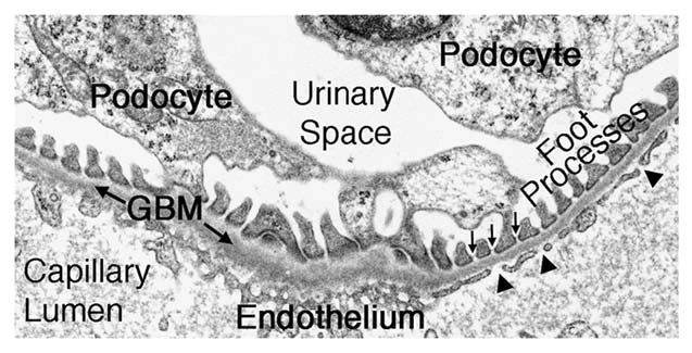
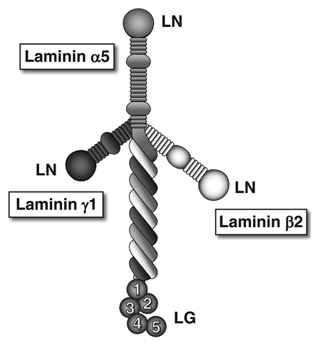
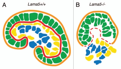
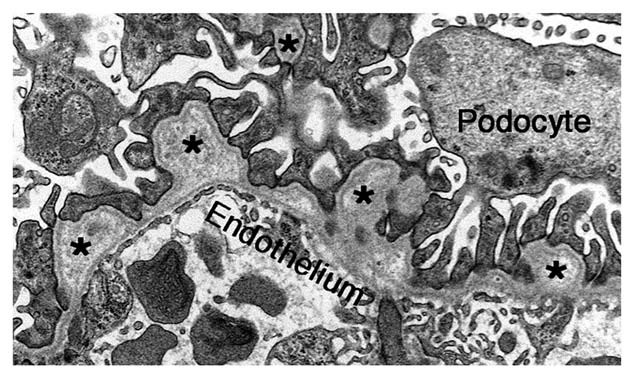
The glomerular basement membrane (GBM) is a crucial component of the kidney's filtration barrier that separates the vasculature from the urinary space. During glomerulogenesis, the GBM is formed from fusion of two distinct basement membranes, one synthesized by the glomerular epithelial cell (podocyte) and the other by the glomerular endothelial cell. The main components of the GBM are laminin-521 (α5β2γ1), collagen α3α4α5(IV), nidogen and the heparan sulfate proteoglycan, agrin. By studying mice lacking specific GBM components, we have shown that during glomerulogenesis, laminin is the only one that is required for GBM integrity and in turn, the GBM is required for completion of glomerulogenesis and glomerular vascularization. In addition, our results from laminin β2-null mice suggest that laminin-521, and thus the GBM, contribute to the establishment and maintenance of the glomerular filtration barrier to plasma albumin. In contrast, mutations that affect GBM collagen IV or agrin do not impair glomerular development or cause immediate leakage of plasma proteins. However, collagen IV mutation, which causes Alport syndrome and ESRD in humans, leads to gradual damage to the GBM that eventually leads to albuminuria and renal failure. These results highlight the importance of the GBM for establishing and maintaining a perfectly functioning, highly selective glomerular filter.


Figures

Figure 1
Figure 1
The ultrastructure of the glomerular filtration barrier. The capillary lumen is lined by an endothelium with fenestrations (arrowheads). The endothelium is adjacent to the ribbon-like glomerular basement membrane (GBM). Podocyte foot processes, which are bridged by slit diaphragms (arrows), abut the opposite aspect of the GBM and are surrounded by the primary filtrate present in the urinary space.
Figure 2
Figure 2
The laminin-521 heterotrimer. This full-sized cruciform laminin is composed of the α5, β2 and γ1 chains. Globular laminin amino-terminal (LN) domains interact with each other to facilitate laminin polymerization in the extracellular matrix. The α chain laminin globular (LG) domain is recognized by receptors on cells, such as integrins, dystroglycan and the Lutheran blood group glycoprotein.
Figure 3
Figure 3
Schematic diagram showing that GBM breakdown due to Lama5 mutation results in failed glomerulogenesis. (A) A maturing wild-type (Lama5+/+) glomerulus with an intact podocyte epithelium (green) and endothelium (yellow) flanking the GBM (red). Mesangial cells (blue) are centrally located within the glomerulus, and parietal epithelial cells (not shown) sit on the Bowman's capsule basement membrane (orange) and line Bowman's space. (B) Failed glomerulogenesis in a Lama5−/− kidney. Discontinuities in the GBM (red) lead to stratification of the podocyte layer (green) and disorganized endothelial and mesangial cells (yellow and blue, respectively).
Figure 4
Figure 4
The ultrastructure of Alport mouse GBM. In the absence of the collagen α3α45α5(IV) network, the GBM becomes split and thickened, with moderately electron-lucent outpocketings (asterisks). However, the endothelium and podocyte foot processes are mostly intact at this early stage of the disease process.

References

    1. Farquhar MG. Editorial: The primary glomerular filtration barrier—basement membrane or epithelial slits? Kidney Int. 1975;8:197–211. - PubMed
    1. St John PL, Abrahamson DR. Glomerular endothelial cells and podocytes jointly synthesize laminin-1 and -11 chains. Kidney Int. 2001;60:1037–1046. - PubMed
    1. Abrahamson DR. Origin of the glomerular basement membrane visualized after in vivo labeling of laminin in newborn rat kidneys. J Cell Biol. 1985;100:1988–2000. - PMC - PubMed
    1. Abrahamson DR, Hudson BG, Stroganova L, Borza DB, St John PL. Cellular origins of type IV collagen networks in developing glomeruli. J Am Soc Nephrol. 2009;20:1471–1479. - PMC - PubMed
    1. Schnabel E, Anderson JM, Farquhar MG. The tight junction protein ZO-1 is concentrated along slit diaphragms of the glomerular epithelium. J Cell Biol. 1990;111:1255–1263. - PMC - PubMed

Publication types

LinkOut - more resources