

Multicystic encephalomalacia as an end-stage finding in abusive head trauma
- PMID: 21519862
- PMCID: PMC3183319
- DOI: 10.1007/s12024-011-9236-7
Multicystic encephalomalacia as an end-stage finding in abusive head trauma
Abstract
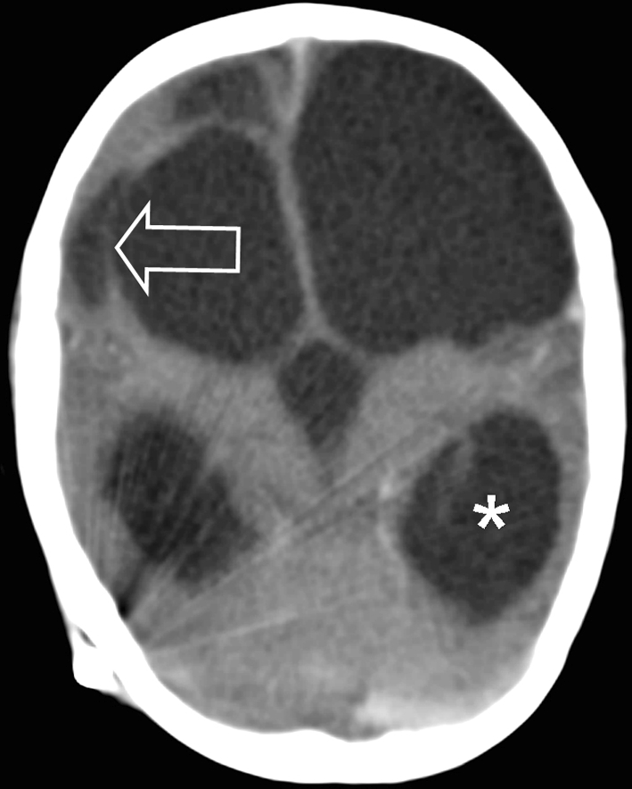
Abusive head trauma (AHT) is one of the most severe forms of physical child abuse. If a child initially survives severe AHT the neurological outcome can be poor. In recent years several children were seen who developed multicystic encephalomalacia (MCE) after documented severe AHT. A search of the Netherlands Forensic Institute database in The Hague was performed. Inclusion criteria were cases of AHT between 1999 and 2010 where the child was under the age of 1 year old at the time of trauma. Trauma mechanism and radiological information were collected. Five children, three boys and two girls (mean age 57 days, range 8-142 days) who developed cystic encephalomalacia after inflicted traumatic brain injury were included. Survival ranged from 27 to 993 days. In all cases judicial autopsy was performed. All cases came before court and in each case child abuse was considered to be proven. In two cases the perpetrator confessed, during police interrogation, to shaking of the child only. Although a known serious outcome, this is one of the few reports on MCE as a result of AHT. In all cases the diagnosis was confirmed at autopsy.
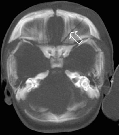
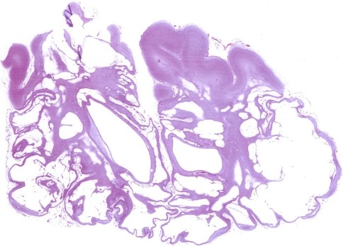
Figures


Similar articles
-
Confessed abusive blunt head trauma.Am J Forensic Med Pathol. 2013 Jun;34(2):130-2. doi: 10.1097/PAF.0b013e31828629ca. Am J Forensic Med Pathol. 2013. PMID: 23629386
-
Abusive head trauma: judicial admissions highlight violent and repetitive shaking.Pediatrics. 2010 Sep;126(3):546-55. doi: 10.1542/peds.2009-3647. Epub 2010 Aug 9. Pediatrics. 2010. PMID: 20696720
-
Long-term outcome in a case of shaken baby syndrome.Med Sci Law. 2016 Apr;56(2):147-9. doi: 10.1177/0025802415581442. Epub 2015 Jun 8. Med Sci Law. 2016. PMID: 26055154
-
Animal models of pediatric abusive head trauma.Childs Nerv Syst. 2022 Dec;38(12):2317-2324. doi: 10.1007/s00381-022-05577-6. Epub 2022 Jun 10. Childs Nerv Syst. 2022. PMID: 35689145 Free PMC article. Review.
-
Aspects of abuse: abusive head trauma.Curr Probl Pediatr Adolesc Health Care. 2015 Mar;45(3):71-9. doi: 10.1016/j.cppeds.2015.02.002. Epub 2015 Mar 12. Curr Probl Pediatr Adolesc Health Care. 2015. PMID: 25771265 Review.
Cited by
-
Parenchymal Brain Laceration as a Predictor of Abusive Head Trauma.AJNR Am J Neuroradiol. 2016 Jan;37(1):163-8. doi: 10.3174/ajnr.A4519. Epub 2015 Oct 15. AJNR Am J Neuroradiol. 2016. PMID: 26471745 Free PMC article.
-
Clinical characteristics of cystic encephalomalacia in children.Front Pediatr. 2024 May 22;12:1280489. doi: 10.3389/fped.2024.1280489. eCollection 2024. Front Pediatr. 2024. PMID: 38840803 Free PMC article.
-
Can Hemorrhagic Stroke Genetics Help Forensic Diagnosis in Pediatric Age (<5 Years Old)?Genes (Basel). 2024 May 13;15(5):618. doi: 10.3390/genes15050618. Genes (Basel). 2024. PMID: 38790247 Free PMC article.
-
What Do Confessions Reveal About Abusive Head Trauma? A Systematic Review.Child Abuse Rev. 2020 May-Jun;29(3):253-268. doi: 10.1002/car.2627. Epub 2020 Jun 23. Child Abuse Rev. 2020. PMID: 37982093 Free PMC article.
-
Functional Recovery in a Patient of Abnormal Left Parieto-Occipital Encephalomalacia With Gliosis-Associated Genu Varum Deformity: A Case Report.Cureus. 2024 Feb 28;16(2):e55115. doi: 10.7759/cureus.55115. eCollection 2024 Feb. Cureus. 2024. PMID: 38558677 Free PMC article.
References
Publication types
MeSH terms
LinkOut - more resources
Full Text Sources
