. 2011 May;53(5):1618-28.
doi: 10.1002/hep.24239.

Ron receptor regulates Kupffer cell-dependent cytokine production and hepatocyte survival following endotoxin exposure in mice

Affiliations

Ron receptor regulates Kupffer cell-dependent cytokine production and hepatocyte survival following endotoxin exposure in mice

William D Stuart et al. Hepatology. 2011 May.

Abstract

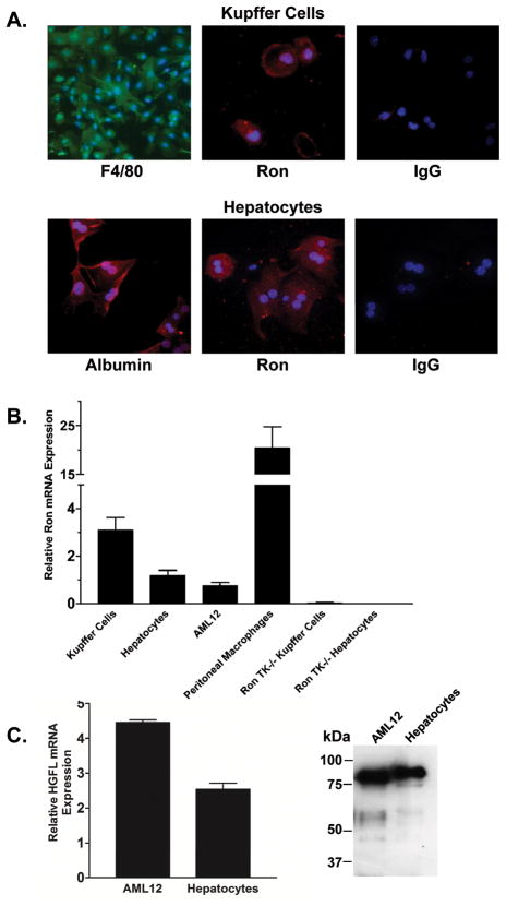
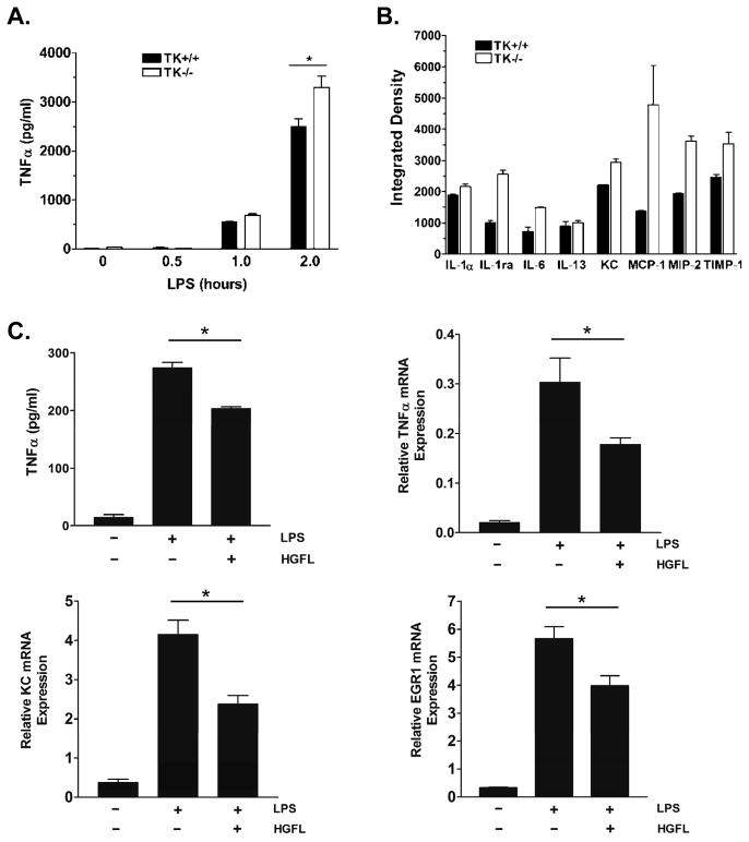
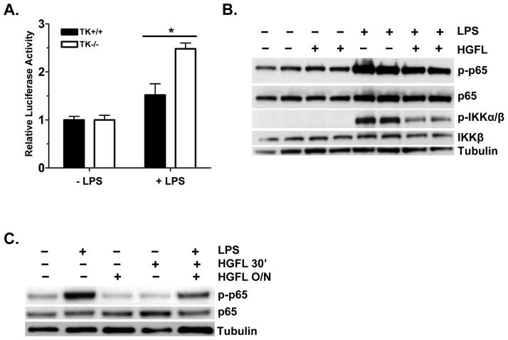
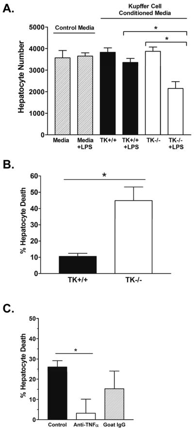
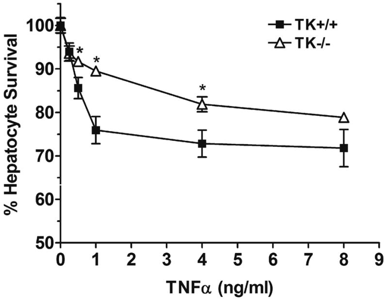
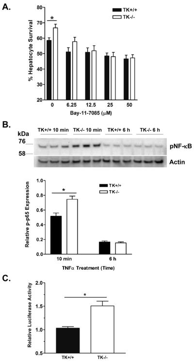
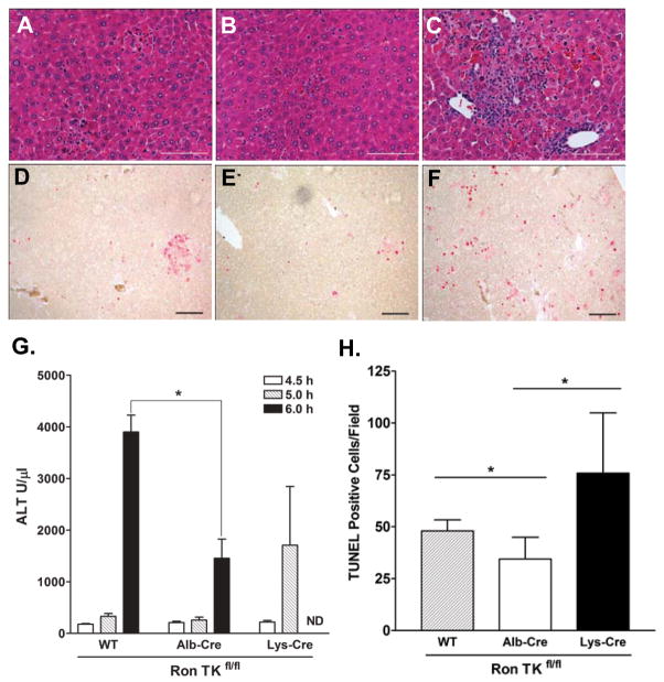
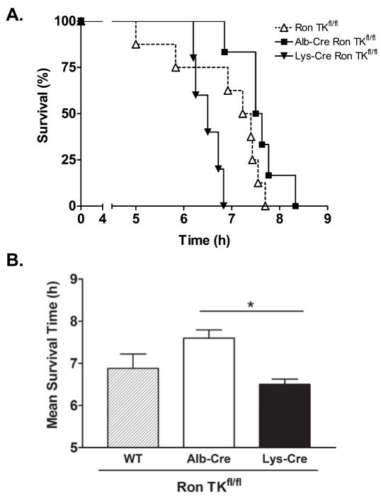
Previous studies demonstrated that targeted deletion of the Ron receptor tyrosine kinase (TK) domain in mice leads to marked hepatocyte protection in a well-characterized model of lipopolysaccharide (LPS)-induced acute liver failure in D-galactosamine (GalN)-sensitized mice. Hepatocyte protection in TK-/- mice was observed despite paradoxically elevated serum levels of tumor necrosis factor alpha (TNF-α). To understand the role of Ron in the liver, purified populations of Kupffer cells and hepatocytes from wildtype (TK+/+) and TK-/- mice were studied. Utilizing quantitative reverse-transcription polymerase chain reaction (RT-PCR), we demonstrated that Ron is expressed in these cell types. Moreover, we also recapitulated the protected hepatocyte phenotype and exaggerated cytokine production observed in the TK-/- mice in vivo through the use of purified cultured cells ex vivo. We show that isolated TK-/- Kupffer cells produce increased levels of TNF-α and select cytokines compared to TK+/+ cells following LPS stimulation. We also show that conditioned media from LPS-treated TK-/- Kupffer cells was more toxic to hepatocytes than control media, suggesting the exaggerated levels of cytokines produced from the TK-/- Kupffer cells are detrimental to wildtype hepatocytes. In addition, we observed that TK-/- hepatocytes were more resistant to cell death compared to TK+/+ hepatocytes, suggesting that Ron functions in both the epithelial and inflammatory cell compartments to regulate acute liver injury. These findings were confirmed in vivo in mice with hepatocyte and macrophage cell-type-specific conditional Ron deletions. Mice with Ron loss selectively in hepatocytes exhibited less liver damage and increased survival compared to mice with Ron loss in macrophages.

Conclusion: We dissected cell-type-specific roles for Ron such that this receptor modulates cytokine production from Kupffer cells and inhibits hepatocyte survival in response to injury.


Figures

Figure 1
Figure 1. Ron is expressed in Kupffer cells and hepatocytes
(A) Representative pictures of isolated Kupffer cells and hepatocytes. Top, Immunocytochemical staining of Kupffer cells by immunofluorescence for the macrophage specific surface marker F4/80 or for Ron compared to control staining with an isotype-matched primary antibody. Nuclei were stained with DAPI. Bottom, Representative pictures depicting Albumin and Ron staining of hepatocytes with DAPI stained nuclei. Control staining with an isotype-matched primary antibody on hepatocytes is shown. (B) Quantitative real-time PCR analysis of Ron mRNA expression from Kupffer cells, hepatocytes, AML12 cells and peritoneal macrophages. Normalized relative Ron expression levels are expressed as the mean ± the standard error and are representative of two independent experiments. Ron expression levels are relative to β-glucuronidase as an internal control. TK−/− Kupffer cells and hepatocytes serve as a negative control. (C) HGFL is expressed at the mRNA level and is secreted by AML12 cells and primary mouse hepatocytes.
Figure 2
Figure 2. Ron regulates cytokine production in Kupffer cells
(A) TNFα ELISA of Kupffer cell media after stimulation with LPS for the indicated times. TNFα production increases in TK+/+ and TK−/− Kupffer cells following LPS stimulation with significantly higher levels in the TK−/− media by 2 hours. *P<0.05. (B) Densitometric analysis of an antibody-based cytokine array of select cytokines from TK+/+ and TK−/− Kupffer cells following LPS stimulation for 2h. Error bars are the range of two spots of each cytokine and is representative of a least two experiments. (C) TNFα ELISA and quantitative real-time PCR of TNFα, KC, and EGR1 showing reduced expression of NF-κB target genes in LPS-stimulated Kupffer cells after overnight treatment with 100 ng/ml HGFL. *P<0.05.
Figure 3
Figure 3. TK−/− Kupffer cells exhibit exaggerated NF-κB signaling in response to LPS
(A) Kupffer cells from TK+/+ and TK−/− mice were transfected with a vector containing an NF-κB response element upstream of firefly luciferase and a control plasmid. After LPS stimulation for 4h, TK−/− Kupffer cells had dramatically higher reporter activity than TK+/+ cells. Bars represent the average of 3 independent experiments with at least two mice per genotype per experiment. *P<0.002. (B) Differentiated THP-1 cells were stimulated with 500 ng/ml of LPS with or without overnight HGFL treatment. Western analysis of cell lysates depicts diminished NF-κB and IKKα/β phosphorylation in HGFL treated cells. Samples from two independent experiments are shown. Total NF-κB, IKKβ, and Tubulin serve as loading controls. (C) Western analysis of LPS-stimulated Kupffer cell lysates with or without HGFL pretreatment overnight (O/N) or for 30 minutes (30′). HGFL treatment before LPS stimulation leads to reduced NF-κB Ser536 phosphorylation (p-p65) compared to controls. Total NF-κB and Tubulin serve as loading controls.
Figure 4
Figure 4. Conditioned media from LPS-stimulated TK−/− Kupffer cells is detrimental to hepatocytes
(A) Kupffer cells from TK+/+ or TK−/− mice were treated in the presence or absence of LPS for 2 hours. Conditioned media was collected and applied to AML12 hepatocytes with ActD. After 18h, the hepatocytes were fixed and quantified by crystal violet staining. (B) Media from the TK−/− Kupffer cells increased hepatocyte death by 35% compared to control cells. The data is expressed as the mean ± standard error and is representative of three independent experiments. *P<0.05. (C) Treatment of TK−/− Kupffer cell conditioned media with an anti-TNFα antibody for 30 minutes leads to a significant reduction in hepatocyte death to less than 5%. Goat IgG was used as a control. Bar graph is the average of 3 experiments. *P<0.05.
Figure 5
Figure 5. TK−/− hepatocytes are protected from TNFα-induced cell death
Primary hepatocytes from TK+/+ or TK−/− mice were treated for 30 minutes with ActD followed by the addition of increasing amounts of TNFα. After 18 hours, the hepatocytes were fixed and cell survival was determined by crystal violet staining with cell number quantitated based on a standard curve of hepatocytes. The data is expressed as the mean ± standard error and is representative of three independent experiments. *P<0.01 compared to the corresponding control treated sample.
Figure 6
Figure 6. NF-κB inhibition abrogates survival of TK−/− hepatocytes in response to TNFα-induced death
(A) Hepatocytes from TK+/+ or TK−/− mice were treated for 30 minutes with ActD plus increasing amounts of the NF-κB activation inhibitor, Bay 11-7085 followed by 5ng/ml TNFα. After 18 hours, the hepatocytes were fixed and cell survival was determined by crystal violet staining. The data is expressed as the mean ± standard error. *P<0.01 (B) Hepatocytes from TK+/+ or TK−/− mice were treated for 30 minutes with ActD followed by 5 ng/ml TNFα. Cells from 3 independent experiments of each cell type were examined after 10 minutes and 6 hours by Western analysis. pNF-κB (phosphorylated p65 subunit at Ser536) band density was normalized to Actin. *P<0.05. (C) TK+/+ and TK−/− hepatocytes were transfected with a vector containing an NF-κB response element upstream of firefly luciferase and a control plasmid. After 10 ng/ml TNFα stimulation for 6h, TK−/− hepatocytes had 1.5 fold higher reporter activity than TK+/+ cells. Values represent the average of two independent experiments relative to untreated cells. *P<0.05.
Figure 7
Figure 7. Deletion of the Ron TK domain in hepatocytes is protective whereas deletion in myeloid derived cells is detrimental to mice treated with LPS/GalN
Histopathology of LPS/GalN mice after 5 hours: (A) Ron TKfl/fl, (B) Alb-Cre Ron TKfl/fl, (C) Lys-Cre Ron TKfl/fl. (D, E, F) Representative TUNEL stained slides (bar=100 μm), positive nuclei stained red. (D) Ron TKfl/fl, (E) Alb-Cre Ron TKfl/fl, (F) Lys-Cre Ron TKfl/fl. (G) Plasma ALT levels of LPS/GalN treated mice at 4.5, 5, and 6 hours. At 6 hours, Ron TKfl/fl control mice had significantly higher ALT than Alb-Cre Ron TKfl/fl mice (*P<0.05). ND, not determined as Lys-Cre mice were moribund or dead at 6 hours. (H) Positive TUNEL counts per 100X field. *P<0.05
Figure 8
Figure 8. Alb-Cre Ron TKfl/fl and Ron TKfl/fl mice have a survival advantage over Lys-Cre Ron TKfl/fl mice treated with LPS/GalN
(A) Kaplan-Meier survival graph of mice injected with LPS/GalN. Lys-Cre Ron TKfl/fl n=5, Ron TKfl/fl n=8, Alb-Cre Ron TKfl/fl n=6. Survival of the Lys-Cre Ron TKfl/fl mice was significantly less than the other two groups. Logrank test P=0.005. (B) Comparison of survival times in hours. *P<0.05 between Alb-Cre and Lys-Cre mice.

References

    1. Ostapowicz G, Fontana RJ, Schiodt FV, Larson A, Davern TJ, Han SH, McCashland TM, et al. Results of a prospective study of acute liver failure at 17 tertiary care centers in the United States. Ann Intern Med. 2002;137:947–954. - PubMed
    1. Bohlinger I, Leist M, Gantner F, Angermuller S, Tiegs G, Wendel A. DNA fragmentation in mouse organs during endotoxic shock. Am J Pathol. 1996;149:1381–1393. - PMC - PubMed
    1. Galanos C, Freudenberg MA. Mechanisms of endotoxin shock and endotoxin hypersensitivity. Immunobiology. 1993;187:346–356. - PubMed
    1. Josephs MD, Bahjat FR, Fukuzuka K, Ksontini R, Solorzano CC, Edwards CK, 3rd, Tannahill CL, et al. Lipopolysaccharide and D-galactosamine-induced hepatic injury is mediated by TNF-alpha and not by Fas ligand. Am J Physiol Regul Integr Comp Physiol. 2000;278:R1196–1201. - PubMed
    1. Wajant H, Pfizenmaier K, Scheurich P. Tumor necrosis factor signaling. Cell Death Differ. 2003;10:45–65. - PubMed

Publication types