. 2011 May;69(5):743-58.
doi: 10.1002/ana.22419.

Neuronal cell death in neonatal hypoxia-ischemia

Affiliations

Neuronal cell death in neonatal hypoxia-ischemia

Frances J Northington et al. Ann Neurol. 2011 May.

Abstract

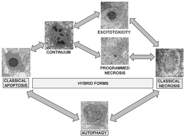
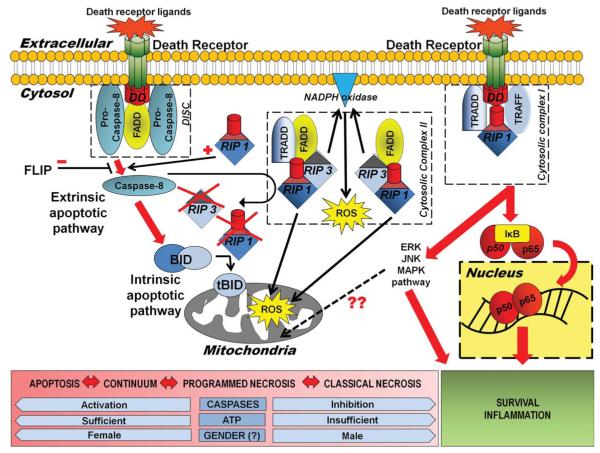
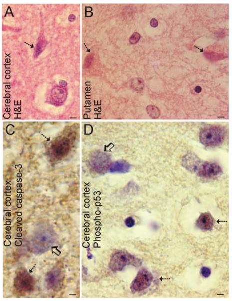
Perinatal hypoxic-ischemic encephalopathy (HIE) is a significant cause of mortality and morbidity in infants and young children. Therapeutic opportunities are very limited for neonatal and pediatric HIE. Specific neural systems and populations of cells are selectively vulnerable in HIE; however, the mechanisms of degeneration are unresolved. These mechanisms involve oxidative stress, excitotoxicity, inflammation, and the activation of several different cell death pathways. Decades ago the structural and mechanistic basis of the cellular degeneration in HIE was thought to be necrosis. Subsequently, largely due to advances in cell biology and to experimental animal studies, emphasis has been switched to apoptosis or autophagy mediated by programmed cell death (PCD) mechanisms as important forms of degeneration in HIE. We have conceptualized based on morphological and biochemical data that this degeneration is better classified according to an apoptosis-necrosis cell death continuum and that programmed cell necrosis has prominent contribution in the neurodegeneration of HIE in animal models. It is likely that neonatal HIE evolves through many cell death chreodes influenced by the dynamic injury landscape. The relevant injury mechanisms remain to be determined in human neonatal HIE, though preliminary work suggests a complexity in the cell death mechanisms greater than that anticipated from experimental animal models. The accurate identification of the various cell death chreodes and their mechanisms unfolding within the immature brain matrix could provide fresh insight for developing meaningful therapies for neonatal and pediatric HIE.


Figures

FIGURE 1
FIGURE 1
Cell death phenotypes in experimental neonatal HI brain injury. After its initial description by Portera-Cailliau and colleagues, the continuum concept, in its original form organized cell death as a linear spectrum with apoptosis and necrosis at the extremes and different syncretic hybrid forms in between. Subsequently, we have found that this concept is fully realized in the neonatal rodent brain following HI, with classically apoptotic and classically necrotic cells found intermixed with various hybrid forms. Cells at the extremes have the well-described structures of necrosis (swelling and vacuolation of organelles, loss of cell membrane integrity, maintenance of nuclear membrane integrity, and random digestion of chromatin) and apoptosis (condensation and darkening of cytoplasm within intact cell membrane, intact organelles until late phases of apoptosis, compaction of chromatin into few uniformly dense and rounded aggregates, and loss of nuclear membrane). Cells with irregular chromatin condensation organized in a “clockface” pattern around an intact nuclear membrane may or may not have preservation of the cytoplasmic membrane and their cytoplasmic organelles are disrupted. This structure has commonly been reported in models of excitotoxicity. Increasing organization of chromatin into regular crescentic or rounded aggregates, partial dissolution of the nuclear membrane and preservation or the cytoplasmic membrane with or without swelling of cytoplasmic organelles within a condensed cytoplasm is the rule, as cell death forms more closely mimic apoptosis and is most commonly referred to as the continuum cell death phenotype. A more recently described cell death phenotype with structural similarity to classical necrosis is referred to as “programmed necrosis” and occurs with random chromatin dissolution, swelling and vacuolation of cytoplasmic organelles including the endoplasmic reticulum and mitochondria as in this example from the neonatal murine model of HI. Various relationships between the classical and hybrid forms of cell death are suggested by their appearance on electron microscopy as represented by the arrows in the figure. Autophagocytic-appearing neurons with large numbers of cytoplasmic vacuoles, partially condensed nuclear chromatin, and preservation of cellular integrity are also found after neonatal HI. Autophagocytic cell death and apoptosis and possibly necrosis also exist on a continuum. Drugs known to inhibit or promote autophagy clearly modulate cell death along the apoptosis-necrosis continuum. HI = hypoxia-ischemia.
FIGURE 2
FIGURE 2
Death receptor signaling and programmed necrosis. This diagram summarizes the possible pleiotropic outcomes following death receptor activation and the signaling pathways leading to these outcomes. Ligand binding to and trimerization of death domain containing members of the TNF receptor superfamily, recruits FADD, a death effector domain containing adaptor protein and procaspase 8, forming the DISC. Signaling for apoptosis then proceeds via the extrinsic or intrinsic pathway. In the extrinsic pathway, active caspase-8 directly cleaves caspase-3. Activation of the mitochondrial or intrinsic pathway proceeds via caspase-8 mediated cleavage of cytosolic Bid. The truncated form of Bid then translocates to mitochondria, thereby functioning as a BH3-only transducer of the death receptor signal at the cell plasma membrane to mitochondria. Simultaneously, cleaved caspase-8 may inactivate death receptor signaling via RIP1 kinase, by cleaving and inactivating both RIP1 and RIP3. Conversely, RIP1 may act to stimulate TNF apoptosis signaling by promoting activation of caspase-8. In the setting of caspase inhibition or severe energy failure, death receptor signaling may preferentially proceed via RIP1 kinase, a death domain containing protein, that binds the death receptor signaling complex containing the adaptor proteins TRADD and TRAF2 at the cell membrane and then TRADD and FADD and RIP3 as cytosolic complex II. Along with RIP3, RIP 1 kinase, death receptor–mediated cell death occurs with ROS production and a cell death morphology resembling necrosis. Both mitochondrial and NADPH oxidase may be the source of enhanced free radical production in this signaling paradigm. When ubiquitinated and bound to the membrane cytosolic complex I, RIP1 kinase activates downstream kinase pathways, which may also contribute to ROS production (possibly via JNK) and also cleave the IKK subunit from the NF-jB complex, initiating NF-κB proinflammatory/prosurvival signaling. Gender likely also has a determinant effect on cell death phenotype following HI. DISC = “death-induced signaling complex”; FADD = Fas-associated death domain; HI = hypoxia-ischemia; IKK = IkappaB kinase complex; JNK = c-Jun N-terminal kinase; NADPH = nicotinamide adenine dinucleotide phosphate; NF-κB = nuclear factor kappa B; RIP1 = receptor interacting protein 1; RIP3 = receptor interacting protein 3; ROS = reactive oxygen species; TNF = tumor necrosis factor; TRADD = TNF receptor-associated death domain; TRAF2 = TNF receptor-associated factor 2.
FIGURE 3
FIGURE 3
Neuronal cell death in human newborn HIE. (A,B) Hematoxylin staining of neocortex from an infant that survived 3 days after HI due to delivery complications reveals selective degeneration of neurons (hatched arrows) in the form of typical ischemic neuronal death with eosinophilic cytoplasm, shrunken cell body, and condensed nucleus. Other damaged neurons are swollen with a vacuolated cytoplasm (hatched arrow in B). This pattern of neurodegeneration is much less phenotypically heterogeneous than that seen in neonatal rodent models of HI but similar to that seen in newborn piglet HI. (A) Bars = 33μm; (B) Bars = 7μm. (C) Subsets of neocortical neurons (hatched arrows) in human infants with HIE display cleaved caspase-3 throughout the cell. Other cells in the microscopic field visualized by the cresyl violet counterstaining have no labeling for cleaved caspase-3 (open arrow). Bar = 15μm. (D) Subsets of neocortical neurons (hatched arrows) in human infants with HIE display active p53 within the nucleus. Other cells (open arrow) in the field have no labeling for active p53. Bar = 15μm. HI = hypoxia-ischemia; HIE = hypoxic-ischemic encephalopathy.

References

    1. Folkerth RD. Neuropathologic substrate of cerebral palsy. J Child Neurol. 2005;20:940–949. - PubMed
    1. Towfighi J, Zec N, Yager J, et al. Temporal evolution of neuropathologic changes in an immature rat model of cerebral hypoxia: a light microscopic study. Acta Neuropathol. 1995;90:375–386. - PubMed
    1. Myers RE. Four patterns of perinatal brain damage and their conditions of occurrence in primates. Adv Neurol. 1975;10:223–234. - PubMed
    1. Adamsons K, Myers RE. Perinatal asphyxia, causes, detection and neurologic sequelae. Pediatr Clin North Am. 1973;20:465–480. - PubMed
    1. Carloni S, Carnevali A, Cimino M, Balduini W. Extended role of necrotic cell death after hypoxia-ischemia-induced neurodegeneration in the neonatal rat. Neurobiol Dis. 2007;27:354–361. - PubMed

Publication types

MeSH terms