

Self-assembly and mineralization of genetically modifiable biological nanofibers driven by β-structure formation
- PMID: 21520924
- PMCID: PMC3114096
- DOI: 10.1021/bm200274r
Self-assembly and mineralization of genetically modifiable biological nanofibers driven by β-structure formation
Abstract
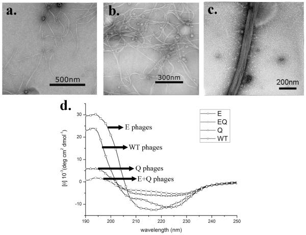
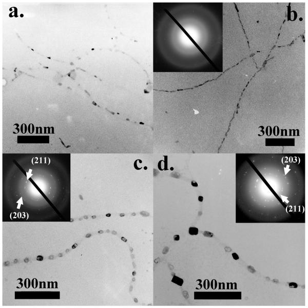
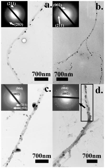
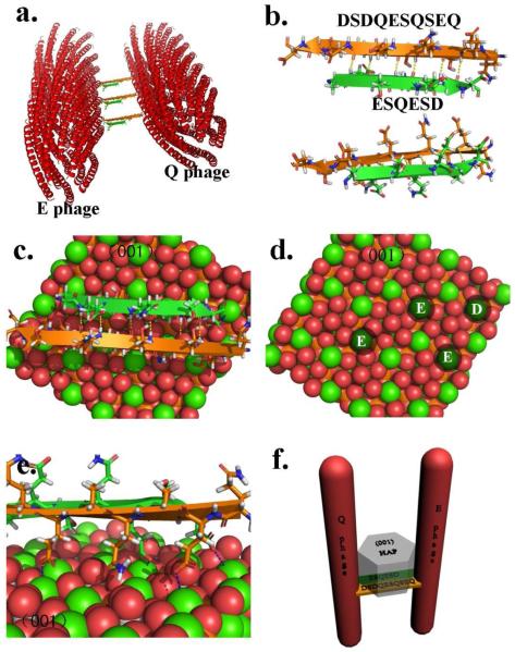
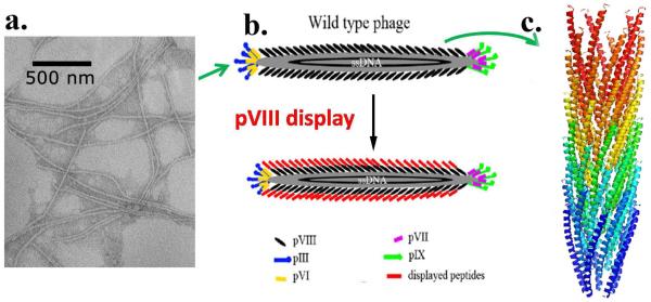
Bioinspired mineralization is an innovative approach to the fabrication of bone biomaterials mimicking the natural bone. Bone mineral hydroxylapatite (HAP) is preferentially oriented with c-axis parallel to collagen fibers in natural bone. However, such orientation control is not easy to achieve in artificial bone biomaterials. To overcome the lack of such orientation control, we fabricated a phage-HAP composite by genetically engineering M13 phage, a nontoxic bionanofiber, with two HAP-nucleating peptides derived from one of the noncollagenous proteins, Dentin Matrix Protein-1 (DMP1). The phage is a biological nanofiber that can be mass produced by infecting bacteria and is nontoxic to human beings. The resultant HAP-nucleating phages are able to self-assemble into bundles by forming β-structure between the peptides displayed on their side walls. The β-structure further promotes the oriented nucleation and growth of HAP crystals within the nanofibrous phage bundles with their c-axis preferentially parallel to the bundles. We proposed that the preferred orientation resulted from the stereochemical matching between the negatively charged amino acid residues within the β-structure and the positively charged calcium ions on the (001) plane of HAP crystals. The self-assembly and mineralization driven by the β-structure formation represent a new route for fabricating mineralized fibers that can serve as building blocks in forming bone repair biomaterials and mimic the basic structure of natural bones.
Figures
References
-
- Mann S. Molecular Recognition in Biomineralization. Nature. 1988;332(6160):119–124.
-
- Berman A, Ahn DJ, Lio A, Salmeron M, Reichert A, Charych D. Total Alignment of Calcite at Acidic Polydiacetylene Films - Cooperativity at the Organic-Inorganic Interface. Science. 1995;269(5223):515–518. - PubMed
-
- Roach H. Why does bone matrix contain non-collagenous proteins? The possible roles of osteocalcin, osteonectin, osteopontin and bone sialoprotein in bone mineralisation and resorption. Cell Biol. Int. 1994;18:617–628. - PubMed
-
- Boskey AL. Matrix proteins and mineralization: an overview. Connect. Tissue Res. 1996;35:357–363. - PubMed
Publication types
MeSH terms
Substances
Grants and funding
LinkOut - more resources
Full Text Sources
Research Materials
