Review
. 2011 Nov;35(6):1126-57.
doi: 10.1111/j.1574-6976.2011.00276.x. Epub 2011 May 31.

Lipoarabinomannan and related glycoconjugates: structure, biogenesis and role in Mycobacterium tuberculosis physiology and host-pathogen interaction

Affiliations
Free PMC article
Review

Lipoarabinomannan and related glycoconjugates: structure, biogenesis and role in Mycobacterium tuberculosis physiology and host-pathogen interaction

Arun K Mishra et al. FEMS Microbiol Rev. 2011 Nov.
Free PMC article

Abstract

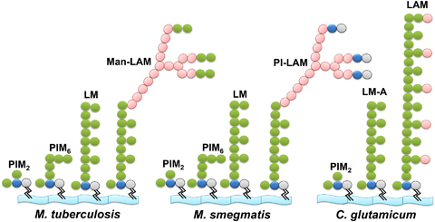
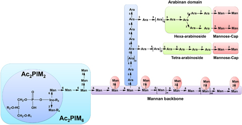
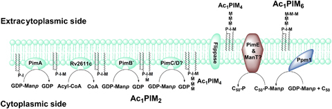
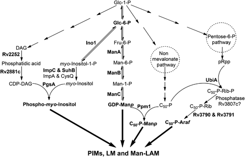
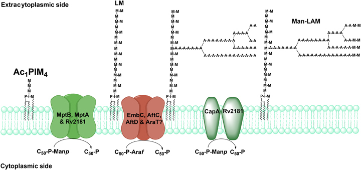
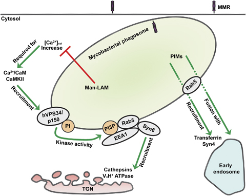
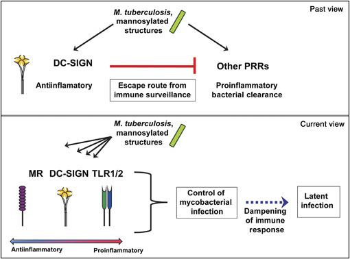
Approximately one third of the world's population is infected with Mycobacterium tuberculosis, the causative agent of tuberculosis. This bacterium has an unusual lipid-rich cell wall containing a vast repertoire of antigens, providing a hydrophobic impermeable barrier against chemical drugs, thus representing an attractive target for vaccine and drug development. Apart from the mycolyl-arabinogalactan-peptidoglycan complex, mycobacteria possess several immunomodulatory constituents, notably lipomannan and lipoarabinomannan. The availability of whole-genome sequences of M. tuberculosis and related bacilli over the past decade has led to the identification and functional characterization of various enzymes and the potential drug targets involved in the biosynthesis of these glycoconjugates. Both lipomannan and lipoarabinomannan possess highly variable chemical structures, which interact with different receptors of the immune system during host-pathogen interactions, such as Toll-like receptors-2 and C-type lectins. Recently, the availability of mutants defective in the synthesis of these glycoconjugates in mycobacteria and the closely related bacterium, Corynebacterium glutamicum, has paved the way for host-pathogen interaction studies, as well as, providing attenuated strains of mycobacteria for the development of new vaccine candidates. This review provides a comprehensive account of the structure, biosynthesis and immunomodulatory properties of these important glycoconjugates.


Figures

Fig. 1
Fig. 1
Lipoarabinomannan and related glycoconjugates found on the cell wall of Mycobacterium tuberculosis, Mycobacterium smegmatis and Corynebacterium glutamicum. Biochemical analysis of the mycobacterial cell wall suggests that different acylated variants of di- and hexa-mannosylated PIMs, Ac1/Ac2PIM2 and PIM6, and the higher glycosylated polymers lipomannan and lipoarabinomannan accumulate in the cell wall. However, in C. glutamicum, only PIM2, two types of lipomannan (LM-A and LM-B, Tatituri et al., 2007a; Mishra et al., 2008b;) and singular Araf capped lipoarabinomannan are present on the cell wall. For the purpose of simplicity, only diacylated forms of these glycoconjugates and LM-A, i.e. MPI anchored lipomannan, are shown. In these glycoconjugates, phosphatidyl-myo-inositol (phosphate in gray and inositol in blue) acts as an anchor to the plasma membrane and further glycosylated by Manp (green) and Araf (pink) sugars yielding different forms of PIMs, lipomannan and lipoarabinomannan that are species specific. In M. tuberculosis and other pathogenic mycobacteria, lipoarabinomannan is capped by mono, -di or -tri α(1→2)-Manp units, resulting in Man-LAM, while in nonpathogenic M. smegmatis, lipoarabinomannan is terminated by phospho inositol, yielding PI-LAM.
Fig. 2
Fig. 2
Schematic structures of lipoarabinomannan and related glycoconjugates. As described in the text, PI acts as an anchor around which PIMs, lipomannan and lipoarabinomannan are built. PI is glycosylated at the 2-OH and 6-OH positions of inositol by Manp residues, and acylated at position 3 of myo-inositol and position 6 of the Manp unit linked at O-2 of myo-inositol in Ac2PIM2 (See the inset in light blue color). Manp at the 6-OH position of inositol is linked to further three and two residues of α(1→6)-Manp and α(1→2)-Manp, respectively, in Ac2PIM6 (see the inset in light indigo color). In lipomannan and the mannan backbone of lipoarabinomannan, PIM2 is linked to another 17–19 residues of Manp in the α(1→6) direction and 7–9 singular branched α(1→2)-Manp units. Mature lipomannan is further linked via an unknown linkage to an arabinan domain made up of approximately 70 Araf residues. The majority of the arabinan domain consists of a linear α(1→5)-Araf polymer branched at certain positions, with α(3→5)-Araf residues towards its nonreducing end resulting in a linear tetra-arabinoside or/and branched hexa-arabinoside domain, which in turn is terminated by β(1→2)-Araf and capped by α(1→2)-Manp units. Here R1, R2, R3 and R4 show different acyl groups found at different locations in the MPI anchor, and n, m, o, p and q represent different degrees of species-specific glycosylation in lipomannan and lipoarabinomannan.
Fig. 4
Fig. 4
Overview of PIM biosynthesis in Mycobacterium tuberculosis. On the cytosolic side of the plasma membrane, PI is glycosylated by PimA, PimB' and an acyltransferase to form Ac1PIM2, which is further mannosylated by PimC and/or PimD? to form Ac1PIM4, an intermediate in Ac1PIM6 and lipomannan biosynthesis. Ac1PIM4 is probably transported across the plasma membrane by unidentified flippases and further mannosylated by α(1→2) mannopyranosyltransferases, PimE and/or another unidentified enzyme to form Ac1PIM6. For simplicity, only triacylated versions of PIMs are shown.
Fig. 3
Fig. 3
Biosynthetic pathways of important nucleotide and lipid-linked sugar donors involved in the synthesis of PIMs, lipomannan and Man-LAM. Most of the sugars utilized by mycobacteria are derived from glycolytic intermediates or glucose as the major carbon source. The experimentally characterized enzymes have been indicated in bold. Apart from the glycolytic pathway, GDP-Manp, C50-P-Manp, C50-P-Araf and PI are also derived from exogenous sources, which have not been shown here to retain simplicity.
Fig. 5
Fig. 5
Biogenesis of Man-LAM from Mycobacterium tuberculosis. Ac1/Ac2PIM4 plausibly serves as an acceptor and extended by MptB in the α(1→6) direction, followed by MptA and further decorated by singular α(1→2)-Manp units by Rv2181 (MptC), resulting in lipomannan. Mature lipomannan is subsequently primed by a singular d-Araf at an unknown position, which is extended by EmbC and/or unidentified α(1→5) arabinofuranosyltransferases. The linear α(1→5)-d-Araf chain is further primed by AftC, which is subsequently extended by AftD and unknown arabinofuranosyltransferases and terminated by the action of AftB to form linear Ara-4 or branched Ara-6. The penultimate Araf of the arabinan domain is further capped by Manp residues by CapA and Rv2181 (MptC) to form Man-LAM. For simplicity, only triacylated versions of different lipoglycans are shown.
Fig. 6
Fig. 6
The role of PIMs and Man-LAM in phagosome maturation arrest by mycobacteria. While Man-LAM prevents lysosomal fusion and acidification, PIMs induce fusion with early endosomes to obtain nutrients required for phagosomal residence of mycobacteria. Man-LAM appears to inhibit cytosolic-Ca2+ increase and thereby blocks the successive steps of hVPS34 kinase activity at the phagosomal membrane, the recruitment of Rab5, EEA1 and Syn6 to the phagosome, and the delivery of cathepsins and VoH+ ATPase. The activity of PIMs in phagosome maturation is dependent on Rab5, but the exact mechanism is not known yet. MMR, macrophage mannose receptor; TGN, trans-Golgi network; CaM, calmodulin; PI, phosphatidylinositol; PI3P, phosphatidylinositol 3-phosphate; Syn, syntaxin; EEA1, early endosome autoantigen 1.
Fig. 7
Fig. 7
The role of DC-SIGN and other PRRs in the immune response against mycobacterial infection. Previously, it was hypothesized that binding to C-type lectin DC-SIGN forms an escape route for mycobacteria to immune surveillance by interfering with signaling via other PRRs. Currently, signaling via PRRs such as DC-SIGN, MMR and TLR2 has been suggested to have a dual function, with both a role in bacterial clearance or induction of proinflammatory cytokines, as well as in preventing an exaggerated immune response. While their main function is then in the protection of the host, Mycobacterium tuberculosis benefits from binding to or signaling via these PRRs as well, which may promote latent mycobacterial infection instead of complete bacterial clearance.

Similar articles

Cited by

References

    1. Akira S, Takeda K. Toll-like receptor signaling. Nat Rev Immunol. 2004;4:499–511. - PubMed
    1. Alderwick LJ, Radmacher E, Seidel M, Gande R, Hitchen PG, Morris HR, Dell A, Sahm H, Eggeling L, Besra GS. Deletion of Cg-emb in Corynebacterineae leads to a novel truncated cell wall arabinogalactan, whereas inactivation of Cg-ubiA results in an arabinan-deficient mutant with a cell wall galactan core. J Biol Chem. 2005;280:32362–32371. - PubMed
    1. Alderwick LJ, Dover LG, Seidel M, Gande R, Sahm H, Eggeling L, Besra GS. Arabinan-deficient mutants of Corynebacterium glutamicum and the consequent flux in decaprenylmonophosphoryl-d-arabinose metabolism. Glycobiology. 2006a;16:1073–1081. - PubMed
    1. Alderwick LJ, Molle V, Kremer L, Cozzone AJ, Dafforn TR, Besra GS, Futterer K. Molecular structure of EmbR, a response element of Ser/Thr kinase signaling in Mycobacterium tuberculosis. P Natl Acad Sci USA. 2006b;103:2558–2563. - PMC - PubMed
    1. Alderwick LJ, Seidel M, Sahm H, Besra GS, Eggeling L. Identification of a novel arabinofuranosyltransferase (AftA) involved in cell wall arabinan biosynthesis in Mycobacterium tuberculosis. J Biol Chem. 2006c;281:15653–15661. - PubMed

MeSH terms