Review
. 2011 Nov;17(8):745-52.
doi: 10.1111/j.1601-0825.2011.01808.x. Epub 2011 Apr 26.

Point-of-care oral-based diagnostics

Affiliations
Review

Point-of-care oral-based diagnostics

R W Hart et al. Oral Dis. 2011 Nov.

Abstract

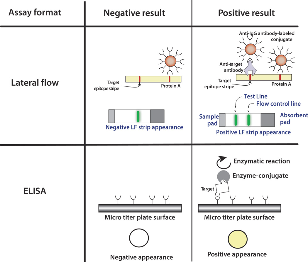
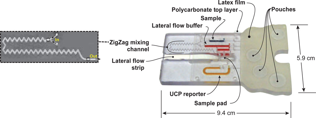
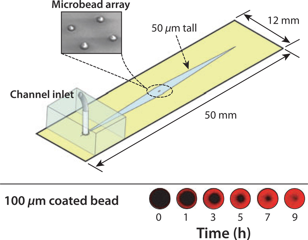
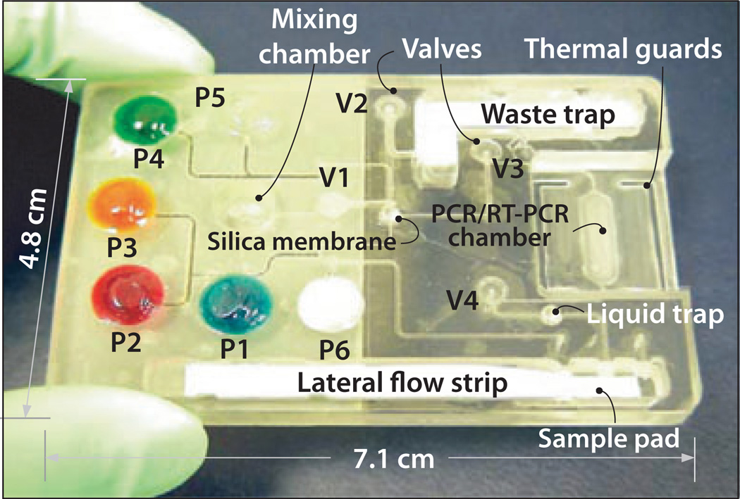
Many of the target molecules that reside in blood are also present in oral fluids, albeit at lower concentrations. Oral fluids are, however, relatively easy and safe to collect without the need for specialized equipment and training. Thus, oral fluids provide convenient samples for medical diagnostics. Recent advances in lab-on-a-chip technologies have made minute, fully integrated diagnostic systems practical for an assortment of point-of-care tests. Such systems can perform either immunoassays or molecular diagnostics outside centralized laboratories within time periods ranging from minutes to an hour. The article briefly reviews recent advances in devices for point-of-care testing with a focus on work that has been carried out by the authors as part of a NIH program.


Figures

Figure 1
Figure 1
A schematic comparing enzyme-linked immunoassay (ELISA) and lateral flow (LF) assays. The ELISA uses an enzyme such as horseradish peroxidase or alkaline phosphatase linked to an antibody to generate either a color change or an electrochemical signal. The ELISA requires multiple washes. The LF assay often uses visual labels such as gold nanoparticles and includes a specific capture line and a control line. The control line binds the labeled conjugate directly without a target and ensures that the sample has flowed up the LF strip
Figure 2
Figure 2
A pouch-based immunoassay cassette. The zigzag conduit (enlarged) facilitates the mixing of the sample with lateral flow (LF) buffer prior to application to the LF strip’s sample pad. The various storage chambers are filled with food coloring for clarity. The assay employs up converting phosphor particles (UCP) as reporters
Figure 3
Figure 3
A schematic depiction of a microbead-based immunoassay chip. The chip contains four porous, agarose beads within a 50-µm tall channel. The beads are sandwiched in place between the floor and ceiling of the channel and remain stationary during continuous flow. The lower image shows the binding of biotin-coated quantum dot labels to the mid-height cross-section of a streptavidin-coated bead as a function of time. The porous structure of the bead and its accessibility to the labels increases the number of binding events that take place in a small volume and improves signal intensity over that obtained with solid beads, which rely solely on peripheral interactions at their surface
Figure 4
Figure 4
A disposable nucleic acid–processing cassette. The plastic cassette hosts a microfluidic network for lysis, nucleic acid isolation by solid-phase extraction, polymerase chain reaction (PCR), and detection of phosphor-labeled PCR products on a lateral flow strip. On-board, fluid-filled pouches are compressed judiciously to propel fluids in the cassette. The actuators of the pouches and valves, the heating, and (optional) real-time detection are housed in a durable analyzer/developer (not shown). The cassette does not exchange any fluids with the analyzer, eliminating the possibility of cross-contamination
Figure 5
Figure 5
Single-chamber chip with an integrated flow through FTA membrane for lysis, nucleic acid isolation, and real-time amplification. Top: Chip’s cross-section. Bottom: chip’s photograph

Similar articles

Cited by

References

    1. Ali MF, Kirby R, Goodey AP, et al. DNA hybridization and discrimination of single-nucleotide mismatches using chip-based microbead arrays. Anal Chem. 2003;75:4732–4739. - PubMed
    1. Ashihara Y, Kasahara Y, Nakamura R. Immunoassay and immunochemistry. Chapter 43. In: McPherson RA, Pincus MR, editors. Henry’s clinical diagnosis and management by laboratory methods. Philadelphia, PA, USA: Saunders Elsevier; 2007. pp. 793–818.
    1. Birkhahn RH, Haines E, Wen W, Reddy L, Briggs WM, Datillo PA. Estimating the clinical impact of bringing a multimarker cardiac panel to the bedside in the ED. [accessed on 5 January 2011];Am J Emerg Med. 2010 Available at: http://www.ncbi.nlm.nih.gov/pubmed/20825823. - PubMed
    1. Blicharz TM, Siqueira WL, Helmerhorst EJ, et al. Fiber-optic microsphere-based antibody array for the analysis of inflammatory cytokines in saliva. Anal Chem. 2009;81:2106–2114. - PMC - PubMed
    1. Bowden M, Song L, Walt DR. Development of a microfluidic platform with an optical imaging microarray capable of attomolar target DNA detection. Anal Chem. 2005;77:5583–5588. - PubMed

Publication types