. 2011 Apr 26;30(1):45.
doi: 10.1186/1756-9966-30-45.

An imbalance in progenitor cell populations reflects tumour progression in breast cancer primary culture models

Affiliations

An imbalance in progenitor cell populations reflects tumour progression in breast cancer primary culture models

Simona Donatello et al. J Exp Clin Cancer Res. .

Abstract

Background: Many factors influence breast cancer progression, including the ability of progenitor cells to sustain or increase net tumour cell numbers. Our aim was to define whether alterations in putative progenitor populations could predict clinicopathological factors of prognostic importance for cancer progression.

Methods: Primary cultures were established from human breast tumour and adjacent non-tumour tissue. Putative progenitor cell populations were isolated based on co-expression or concomitant absence of the epithelial and myoepithelial markers EPCAM and CALLA respectively.

Results: Significant reductions in cellular senescence were observed in tumour versus non-tumour cultures, accompanied by a stepwise increase in proliferation:senescence ratios. A novel correlation between tumour aggressiveness and an imbalance of putative progenitor subpopulations was also observed. Specifically, an increased double-negative (DN) to double-positive (DP) ratio distinguished aggressive tumours of high grade, estrogen receptor-negativity or HER2-positivity. The DN:DP ratio was also higher in malignant MDA-MB-231 cells relative to non-tumorigenic MCF-10A cells. Ultrastructural analysis of the DN subpopulation in an invasive tumour culture revealed enrichment in lipofuscin bodies, markers of ageing or senescent cells.

Conclusions: Our results suggest that an imbalance in tumour progenitor subpopulations imbalances the functional relationship between proliferation and senescence, creating a microenvironment favouring tumour progression.


Figures

Figure 1
Figure 1
Characterization of tumour and non-tumour primary cultures. A. Organoid-derived cultures (A, top panels, 10X magnification) from both tumour and non-tumour specimens had large polygonal cells (lower panels, lpc) surrounded by small polygonal cells (lower panels, spc, 20X magnification). B. Representative tumour and non-tumour cultures (passages 1-3) were analyzed for expression of the epithelial markers K19, K18 and ESA and the myoepithelial markers SMA, K14 and vimentin (scale bar 50 μm). C. Representative cultures were immunoblotted for expression of epithelial (K19, K18) and myoepithelial (vimentin, p63) markers.
Figure 2
Figure 2
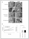
Ultrastructural and functional differences distinguish non-tumour from tumour primary cultures. A. TEM analysis of non-tumour cells revealed modest numbers of cytoplasmic vesicles (Vnt), single nuclei, distinct nuclear double membranes (NMnt), regular mitochondria (Mnt) and well-organized RER (Rnt). Tumour cells showed abnormal peri-nuclear vesicles (Vt), >1 nucleus per cell with thin nuclear membranes (NMt), abnormal mitochondria (Mt) and disorganized RER (Rt). B. Proliferation was enhanced in HG tumour cultures relative to LG tumour cultures or non-tumour cultures (left). Basal senescence, estimated by SA-β-galactosidase staining, was lower in tumour versus non-tumour cultures (right; p < 0.001).
Figure 3
Figure 3
Ultrastructural identification of putative progenitor cells in primary cultures. HMEC and tumour primary cultures analyzed by TEM were observed to grow as multi-layers, with basally-located cells having plump morphologies (a, arrow) compared to the attenuated morphologies of apically-located cells. Filament-rich cells (b, arrows) were layered with glycogen-rich cells (b, arrowhead). A schematic representation of cellular organization is shown in (c).
Figure 4
Figure 4
Isolation of putative progenitor cells from primary cultures and cell lines. A. Breast primary cultures were sorted into CALLA single-positive, EPCAM single-positive, double-positive (DP) or double-negative (DN) populations, and expressed as a percentage of total cells. B. TEM analysis revealed a high content of lipofuscin bodies in the DN population sorted from a tumour culture (arrows). C. The DN:DP ratio increased in three types of aggressive tumour (high grade, ER-negative or HER2-positive) relative to non-tumour or non-aggressive tumour cultures. D. The DN:DP ratio in metastatic MDA-MB-231 cells exceeded that in non-tumourogenic MCF-10A cells. E. Activity of the stem cell marker ALDH was similar in non-tumour versus pooled tumour cultures (left), but significantly higher in non-tumour and low grade tumour cultures compared to high grade tumour cultures (p < 0.001; right).
Figure 5
Figure 5
Progenitor imbalance model. A normal phenotype likely requires a fine balance between different progenitor populations (DP and DN). In normal cells, a balance between proliferation and senescence interplays with a balance between these putative progenitor populations. This promotes regulated generation of differentiated cells. In aggressive tumours, increased proliferation and decreased senescence influences the equilibrium between different progenitor populations. This may alter the differentiated/undifferentiated cell balance, promoting basal-like phenotypes associated with tumour progression.

Similar articles

Cited by

References

    1. Molyneux G, Geyer FC, Magnay FA, McCarthy A, Kendrick H, Natrajan R, Mackay A, Grigoriadis A, Tutt A, Ashworth A, BRCA1 basal-like breast cancers originate from luminal epithelial progenitors and not from basal stem cells. Cell Stem Cell. pp. 403–417. - PubMed
    1. Kakarala M, Wicha MS. Implications of the cancer stem-cell hypothesis for breast cancer prevention and therapy. J Clin Oncol. 2008;26:2813–2820. doi: 10.1200/JCO.2008.16.3931. - DOI - PMC - PubMed
    1. Stingl J, Eaves CJ, Kuusk U, Emerman JT. Phenotypic and functional characterization in vitro of a multipotent epithelial cell present in the normal adult human breast. Differentiation. 1998;63:201–213. doi: 10.1111/j.1432-0436.1998.00201.x. - DOI - PubMed
    1. Clayton H, Titley I, Vivanco M. Growth and differentiation of progenitor/stem cells derived from the human mammary gland. Exp Cell Res. 2004;297:444–460. doi: 10.1016/j.yexcr.2004.03.029. - DOI - PubMed
    1. Ginestier C, Hur MH, Charafe-Jauffret E, Monville F, Dutcher J, Brown M, Jacquemier J, Viens P, Kleer CG, Liu S. et al.ALDH1 is a marker of normal and malignant human mammary stem cells and a predictor of poor clinical outcome. Cell Stem Cell. 2007;1:555–567. doi: 10.1016/j.stem.2007.08.014. - DOI - PMC - PubMed

Publication types