Review
. 2011 Apr 28;70(2):178-99.
doi: 10.1016/j.neuron.2011.04.007.

The expanding social network of ionotropic glutamate receptors: TARPs and other transmembrane auxiliary subunits

Affiliations
Review

The expanding social network of ionotropic glutamate receptors: TARPs and other transmembrane auxiliary subunits

Alexander C Jackson et al. Neuron. .

Abstract

Ionotropic glutamate receptors (iGluRs) underlie rapid, excitatory synaptic signaling throughout the CNS. After years of intense research, our picture of iGluRs has evolved from them being companionless in the postsynaptic membrane to them being the hub of dynamic supramolecular signaling complexes, interacting with an ever-expanding litany of other proteins that regulate their trafficking, scaffolding, stability, signaling, and turnover. In particular, the discovery that transmembrane AMPA receptor regulatory proteins (TARPs) are AMPA receptor auxiliary subunits that are critical determinants of their trafficking, gating, and pharmacology has changed the way we think about iGluR function. Recently, a number of novel transmembrane proteins have been uncovered that may also serve as iGluR auxiliary proteins. Here we review pivotal developments in our understanding of the role of TARPs in AMPA receptor trafficking and gating, and provide an overview of how newly discovered transmembrane proteins expand our view of iGluR function in the CNS.


Figures

FIGURE 1
FIGURE 1. Major structural domains of AMPARs and TARPs
Illustration of the structural features of a closely apposed GluA subunit (left) and a canonical TARP auxiliary subunit (right). The GluA subunit of a tetrameric AMPAR is composed of a large extracellular N-terminal domain (NTD), the ligand-binding core, transmembrane domains, linker regions and several intracellular domains including the C-terminal tail (CTD). Agonists such as glutamate (yellow) bind within the ligand-binding core to mediate channel opening. The Q/R site (magenta) is the narrowest constriction of the AMPAR pore and is an important determinant of its functional properties. The TARP auxiliary subunit consists of four transmembrane domains with a large extracellular loop, which is essential for TARP modulation of AMPAR gating. The tip of the TARP CTD contains a PDZ binding motif (red), which is known to bind to PDZ domain-containing proteins such as PSD-95, and is essential for the synaptic targeting of AMPARs.
FIGURE 2
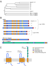
FIGURE 2. The TARP family of transmembrane AMPAR auxiliary subunits and their relatives
(A) Dendrogram illustrating the approximate phylogenetic relationships between all known TARP auxiliary subunits and several related proteins. The TARPs include stargazin (γ-2), γ-3, γ-4, γ-5, γ-7 and γ-8. The TARPs are homologous to the skeletal muscle voltage-gated calcium channel auxiliary subunit, γ-1, as well as γ-6. Claudin-1 is a member of the claudin family of tight-junction proteins. The more distantly related proteins from C. elegans, SOL-1, STG-1 and STG-2 are necessary for the function of GLR-1, the AMPAR homolog in C. elegans. The dendrogram is based on sequence alignment of amino acid sequences using ClustalW. All protein sequences are from mouse unless otherwise noted (C. elegans). (B) Bar diagrams showing the predicted domain structures of the TARPs (both type I and type II), in addition to C. elegans STG-1/2 and SOL-1 for comparison. Noteworthy are the four large extracellular CUB domains of SOL-1 as well as the approximate locations of sites of post-translational modification. (C) Illustration of the proposed secondary structures of the proteins shown in (B). Top is extracellular, bottom is intracellular.
FIGURE 3
FIGURE 3. TARP modulation of AMPAR gating and pharmacology
Schematic summary of the myriad ways in which TARP association can modulate the gating and pharmacology of AMPARs. Note that not every TARP effect on AMPAR function is illustrated here. TARP modulation (red) is shown relative to GluA alone (black). Represented are TARP-dependent changes in deacativation and desensitization kinetics as well as the phenomenon known as resensitization. TARPs are known to modulate other channel properties such as mean channel conductance, open probability and intracellular polyamine affinity. TARPs also modulate AMPAR pharmacology in the form of changes in glutamate affinity, kainate efficacy, CNQX efficacy and sensitivity to polyamine toxins such as philanthotoxin (PhTx). These functional properties vary in a combinatorial manner depending on TARP subtype and AMPAR subunit composition.
FIGURE 4
FIGURE 4. Candidate iGluR transmembrane auxiliary subunits
(A) Bar diagrams showing the predicted domain structures of four candidate iGluR transmembrane auxiliary subunits. CNIH-2/3, CKAMP44 and SynDIG1 have been shown to bind to and influence the trafficking and/or gating of AMPARs. CNIH-2/3 is a small protein with three predicted transmembrane domains. CKAMP44 has a single transmembrane domain, a cysteine-rich extracellular N-terminal domain (likely forming a cystine knot) and a long intracellular CTD tail ending in a PDZ-binding motif. SynDIG1 also has only one predicted transmembrane domain with a C-terminal hydrophobic region. NETO-1 is a candidate NMDAR auxiliary subunit, while NETO-2 is a candidate KAR auxiliary subunit. These homologous proteins are represented by one bar diagram, which highlights the two large extracellular CUB domains and a long CTD ending in a PDZ-binding motif. (B) Illustration of the proposed secondary structures of the proteins shown in (A). Top is extracellular, bottom is intracellular.

References

    1. Abouda H, Hizem Y, Gargouri A, Depienne C, Bouteiller D, Riant F, Tournier-Lasserve E, Gourfinkel-An I, LeGuern E, Gouider R. Familial form of typical childhood absence epilepsy in a consanguineous context. Epilepsia. 2010;51:1889–1893. - PubMed
    1. Arbuzova A, Schmitz AA, Vergères G. Cross-talk unfolded: MARCKS proteins. Biochem. J. 2002;362:1–12. - PMC - PubMed
    1. Arikkath J, Campbell KP. Auxiliary subunits: essential components of the voltage-gated calcium channel complex. Curr. Opin. Neurobiol. 2003;13:298–307. - PubMed
    1. Armstrong N, Sun Y, Chen GQ, Gouaux E. Structure of a glutamate-receptor ligand-binding core in complex with kainate. Nature. 1998;395:913–917. - PubMed
    1. Ascher. P, Nowak L. Quisqualate- and kainate-activated channels in mouse central neurones in culture. J. Physiol. 1988;399:227–245. - PMC - PubMed

Publication types

MeSH terms

LinkOut - more resources