. 2011 Apr 28;70(2):266-80.
doi: 10.1016/j.neuron.2011.03.013.

The oriented emergence of axons from retinal ganglion cells is directed by laminin contact in vivo

Affiliations

The oriented emergence of axons from retinal ganglion cells is directed by laminin contact in vivo

Owen Randlett et al. Neuron. .

Abstract

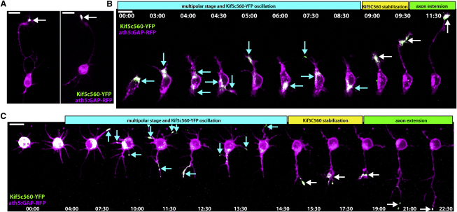
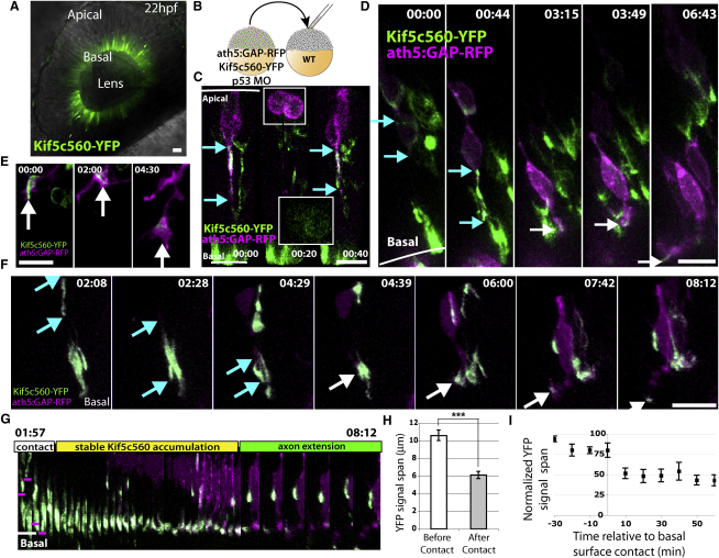
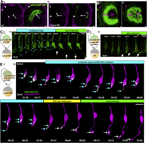
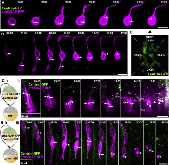
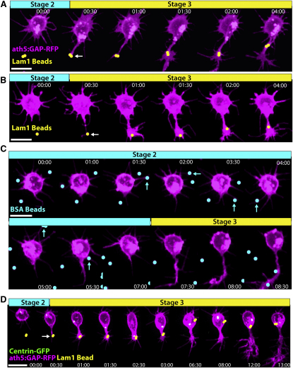
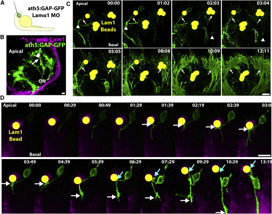
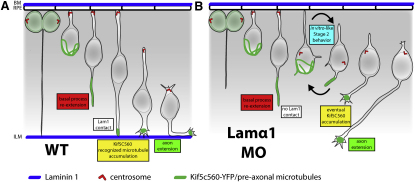
How the site of axon emergence is specified during neural development is not understood. Previous studies disagree on the relative importance of intrinsic and extrinsic mechanisms. The axons of retinal ganglion cells (RGCs) emerge basally in vivo, yet because RGCs develop from polarized neuroepithelial cells within a polarized environment, disentangling intrinsic and extrinsic influences is a challenge. We use time-lapse imaging to demonstrate that Laminin acting directly on RGCs is necessary and sufficient to orient axon emergence in vivo. Laminin contact with the basal processes of newborn RGCs prevents the cells from entering a stochastic Stage 2 phase, directs the rapid accumulation of the early axonal marker Kif5c560-YFP, and leads to the formation of axonal growth cones. These results suggest that contact-mediated cues may be critical for the site of axon emergence and account for the differences in cellular behavior observed in vitro and in vivo.


Figures

Figure 1
Figure 1
Kif5c560-YFP Marks the Axon of Cultured RGCs and Oscillates during Stage 2 Retinal ganglion cells (RGCs) were dissociated from ath5:GAP-RFP embryos injected with Kif5c560-YFP mRNA at the one-cell stage and allowed to polarize in vitro. (A) Bright Kif5c560-YFP was seen in the growth cone of the long axon projecting from polarized RGCs (arrow). (B and C) Shortly after plating, RGCs exhibit Kif5c560-YFP oscillations in different areas of the cell body and individual neurites, typical of Stage 2 behavior (arrows, cyan phase). The construct eventually stabilizes in a single neurite (white arrows, yellow phase), and this neurite extends to form the axon (green phase). Figure is related to Movie S1. Time is shown in hr:min; scale bars = 10 μM.
Figure 2
Figure 2
Kif5c560-YFP Marks the Axon in RGCs In Vivo, and Accumulates in a Highly Directed Manner at the Tip of the Basal Process Prior to Axon Extension (A) Injection of Kif5c560-YFP mRNA at the one-cell stage results in expression in all retinal cells, and demonstrates that Kif5c560 accumulates basally in neuroepithelial cells. Image is a single confocal slice. (B) Transplantation scheme used to create mosaic embryos expressing ath5:GAP-RFP and Kif5c560-YFP in a subset of RGCs. (C) Kif5c560-YFP signal accumulation (marked by cyan arrows) is confined to the basal process of ath5-expressing neuroepithelial cells (t = 0:00) and to young postmitotic neuroblasts (t = 0:40), but is diffuse during mitosis (t = 0:20, inset). (D) In very young RGCs with low ath5:GAP-RFP signal, Kif5c560-YFP signal is seen in the cell body (t = 0:00, YFP span marked by cyan arrows), but quickly moves to the basal process (t = 0:44), to the tip of the basal process (t = 3:15, white arrows), and finally to the extending axonal growth cone (t = 6:43). (E) A higher-magnification example showing an RGC growth cone (arrows) navigating within the retina and demonstrating strong and specific Kif5c560-YFP accumulation. (F) Time-lapse image demonstrating that Kif5c560-YFP signal initially spans a large portion of the basal process (span marked by cyan arrows), but upon contact with the basal surface, YFP signal accumulates specifically at the tip of the basal process (white arrows), and finally in the extending axon. (G) Kymograph of the cell shown in (F), demonstrating that immediately after the RGC basal process (marked by magenta line) contacts the basal surface of the retina (white line), the Kif5c560-YFP signal accumulates specifically at the tip of the basal process before the axon extends. (H) Measuring the length of Kif5c560-YFP signal span demonstrates a significant decrease in length after basal surface contact (mean ± SEM, p < 0.0001, Mann-Whitney test, n = 7 cells from three embryos). (I) Plotting the YFP signal span normalized to the longest observed length for each cell (mean ± SEM) demonstrates that the specific accumulation of YFP occurs immediately after basal surface contact (t = 0). Frames are taken from Movie S2. Time is shown in hr:min; scale bars = 10 μM.
Figure 3
Figure 3
Lamα1 Is Necessary for Directed RGC Polarization (Ai) Immunofluorescent staining of ath5:GAP-GFP embryos with polyclonal rabbit anti-Lam1 antibody reveals strong staining at the basal lamina lining the basal surface of the retina, or ILM (B, arrow), as well as at the basal lamina of the RPE, or Bruch's membrane (A, arrowhead). (Aii) Injection of an antisense morpholino targeted against Lamα1 results in efficient loss of Lam1 staining at the basal surface (arrow), while Lam1 staining at Bruch's membrane remains. Images are of a single confocal slice. (B) Confocal reconstruction from a WT 3 dpf retina (Bi) demonstrating the highly ordered nature of the ganglion cell layer (GCL) and the RGC axon fascicles () collecting to form the optic nerve (ON). (Bii) After lamα1 morpholino injection, the GCL is disorganized, as are the axon fascicles, which meander through the retina before colleting to form the ON. (Ci) Mosaic embryos with WT ath5:GAP-GFP-labeled RGCs in a lamα1 morphant environment were analyzed by time-lapse confocal microscopy beginning at approximately 35 hpf. (Cii) RGCs in this environment progress through a transient multipolar phase (marked by [], cyan phase) before projecting an axon (arrow, green phase). (Di) Mosaic embryos with lamα1 morphant, ath5:GAP-GFP-labeled RGCs in a WT environment were analyzed by time-lapse confocal microscopy. (Dii) Morphant RGCs behave normally in this environment, and axons project directly from the basal surface of the cell (marked by arrowheads). (Ei) Mosaic embryos with WT ath5:GAP-GFP-labeled, Kif5c560-YFP-expressing RGCs in a lamα1 morphant environment were analyzed by time-lapse confocal microscopy. (Eii) In this context, Kif5c560-YFP signal accumulation (marked by cyan arrows) oscillates between the cell body and transient neurites (cyan phase) before stably accumulating in a single neurite (marked by white arrows, yellow phase) that extends to form the axon (green phase). Note that the individual confocal z-slices were cropped to remove signal not associated with the cell. A reconstruction of the uncropped frames is shown in Movie S5. Frames are taken from Movies S3, S4, and S5. Time is shown in hr:min; scale bars = 10 μM.
Figure 4
Figure 4
Centrosomes Are Static and Apically Localized in RGCs Polarizing in WT Retinas, but Mislocalized and Dynamic in RGCs Polarizing In Vitro and in lamα1 Morphants (A and B) RGCs from ath5:GAP-RFP/Centrin-GFP transgenic embryos were dissociated and imaged during polarization in vitro. Centrosomes (marked by arrow) are not stably localized, but instead move dynamically within the cell body and even within Stage 2 neurites. (C) Overlay of the centrosomes from 33 polarized RGCs in vitro showing their position in reference to the base of the axon (arrow, ath5:GAP-RFP channel not shown). Centroid analysis demonstrates that centrosome location is not biased to any specific quadrant of the RGC cell body (p = 0.9536: Chi square test, n = 33 cells). (Di) Mosaic embryos with WT ath5:GAP-RFP/Centrin-GFP-labeled RGCs in a WT environment were analyzed by time-lapse confocal microscopy. (Dii) Centrosomes (marked by arrows) remain apically positioned in RGCs up until dendrite formation and IPL stratification (marked by arrowheads, t = 18:13). (Ei) Mosaic embryos with WT ath5:GAP-RFP/Centrin-GFP-labeled RGCs in a lamα1 morphant environment were analyzed by time-lapse confocal microscopy. (Eii) The centrosome (marked by arrow) is initially localized apically (t < 07:00), but becomes mislocalized during RGC polarization, and moves dynamically within the cell body. Frames are taken from Movies S6 and S7. Time is shown in hr:min; scale bars = 10 μM.
Figure 5
Figure 5
Lam1 Contact Is Sufficient to Transform a Stage 2 Neurite into an Axon In Vitro Dissociated RGCs from ath5:GAP-RFP embryos were plated on poly-L-lysine scattered with Lam1-coated 1 μM polystyrene beads (pseudocolored yellow) and analyzed by time-lapse microscopy. (A and B) When a Stage 2 neurite contacts a Lam1 bead (contact point marked by arrow), this quickly (within one 30 min time point) induces a dramatic transformation from a thin neurite to one with an elaborate growth cone typical of an RGC axon. (C) When presented with BSA-coated control beads (pseudocolored cyan), neurite contact (cyan arrows) does not have an observable effect. (D) Imaging of cultured ath5:GAP-RFP/Centrin-GFP RGCs contacting a Lam1 bead (pseudocolored yellow, arrow indicates contact point) demonstrates that along with axon induction, neurite contact causes the centrosome to orient toward the site of Lam1 contact, and induces a transient migration toward the bead. Frames are taken from Movies S8 and S9. Time is shown in hr:min; scale bars = 10 μM.
Figure 6
Figure 6
Lam1 Contact Is Sufficient to Transform a Neurite into an Axon In Vivo (A) ath5:GAP-GFP transgenic, lamα1 morphant embryos were grown to ∼24 hpf, and Lam1-coated polystyrene beads were implanted into the retina of the right eye. (B) Confocal reconstruction of an embryo that was implanted with a clump of 1 μM Lam1 beads and grown until RGC axons had extended (∼3 dpf), immunostained with polyclonal anti-Lam1 antibody. This demonstrates the intimate association between the highly stained Lam1-coated beads and the extended axons. Axons hug the surface of the bead clump (arrow), causing the beads to lie within the axon fascicle. (C) Time-lapse confocal imaging during axon extension demonstrates the dramatic interaction between polarizing RGC axons and the Lam1-coated beads (pseudocolored yellow), resulting in axon extension along the surface of the beads (visible growth cones marked by arrowheads) and bead engulfment by the axon fascicle (arrows). (D) Highlighting an individual RGC by pseudocoloring it green demonstrates that after Lam1 bead contact (white arrows), the contacting neurite transforms into a process tipped with an elaborate growth cone that extends to form the axon, while the axon shaft remains associated with the bead (blue arrows). Frames are taken from Movies S10 and S12. Time is shown in hr:min; scale bars = 10 μM.
Figure 7
Figure 7
Lam1 Contact Directs Kif5c560-YFP to the Contacting Neurite In Vitro and In Vivo (A–C) RGCs were dissociated from ath5:GAP-RFP embryos injected with Kif5c560-YFP mRNA at the one-cell stage, plated on poly-L-lysine with scattered 1 μM Lam1-coated beads (A and B, pseudocolored yellow), or on islands of Lam1 stained with Texas red dye (C, magenta substrate). When a single neurite contacted Lam1 (A), this quickly caused the YFP signal to accumulate specifically in that neurite (arrow). When multiple neurites contacted Lam1 (B and C), this caused the YFP signal to accumulate specifically in Lam1-contacting neurites, and to oscillate preferentially between these neurites (arrows). (Di) Blastomeres were transplanted from ath5:GAP-RFP transgenic embryos that were injected with Kif5c560-YFP mRNA into lamα1 morphant embryos or mosaic embryos grown to 24 hpf, and 6 μM Lam1-coated beads were implanted into the right eye. (Dii) The highlighted RGC exhibits typical Kif5c560-YFP oscillations during Stage 2 (cyan phase, marked by red arrowheads). Upon contact with the Lam1 bead (white arrowhead; bead is pseudocolored yellow), the contacting process is stabilized and does not retract, and YFP signal is concentrated at the contact point. This neurite then extends to form the axon (green phase). Note that the individual confocal z-slices were cropped to remove signal not associated with the cell. A reconstruction of the uncropped frames is shown in Movie S16. (E) Kymograph of the cell shown in (D), demonstrating the specific Kif5c560-YFP signal accumulation upon neurite contact with the Lam1-coated bead (contact marked by white arrow; bead pseudocolored yellow). Frames are taken from Movies S13, S14, S15, and S16. Time is shown in hr:min; scale bars = 10 μM.
Figure 8
Figure 8
Model for Directed RGC Axon Emergence (A) In a WT retina, the newly born RGC re-extends a basal process toward the basal surface of the retina. The process contacts Lam1 within the retinal basal lamina at the ILM (blue). This stabilizes the basal process, causes Kif5c560-YFP-recognized microtubules (green) to accumulate specifically at the contact point, and commits this neurite to form the axon. (B) In a Lam1-deficient retina, the re-extending basal process does not contact the Lam1 cue, causing it to retract, and causing the cell to enter an ectopic Stage 2 phase typical of neurons polarizing in vitro. During this phase transient neurites are extended, Kif5c560-YFP signal oscillates within the cell, and the centrosome becomes mislocalized and dynamic. Driven by the intrinsic polarization program, and perhaps by other intrinsic/extrinsic cues, Kif5c560-YFP signal eventually stabilizes in a single neurite that matures and extends to form the axon.

References

    1. Adler C.E., Fetter R.D., Bargmann C.I. UNC-6/Netrin induces neuronal asymmetry and defines the site of axon formation. Nat. Neurosci. 2006;9:511–518. - PMC - PubMed
    1. Arimura N., Kaibuchi K. Neuronal polarity: from extracellular signals to intracellular mechanisms. Nat. Rev. Neurosci. 2007;8:194–205. - PubMed
    1. Barnes A.P., Polleux F. Establishment of axon-dendrite polarity in developing neurons. Annu. Rev. Neurosci. 2009;32:347–381. - PMC - PubMed
    1. Barnes A.P., Lilley B.N., Pan Y.A., Plummer L.J., Powell A.W., Raines A.N., Sanes J.R., Polleux F. LKB1 and SAD kinases define a pathway required for the polarization of cortical neurons. Cell. 2007;129:549–563. - PubMed
    1. Basto R., Lau J., Vinogradova T., Gardiol A., Woods C.G., Khodjakov A., Raff J.W. Flies without centrioles. Cell. 2006;125:1375–1386. - PubMed

Publication types

MeSH terms

LinkOut - more resources