. 2011 Apr;23(4):1523-35.
doi: 10.1105/tpc.110.081067. Epub 2011 Apr 26.

Arabidopsis thaliana high-affinity phosphate transporters exhibit multiple levels of posttranslational regulation

Affiliations

Arabidopsis thaliana high-affinity phosphate transporters exhibit multiple levels of posttranslational regulation

Vincent Bayle et al. Plant Cell. 2011 Apr.

Abstract

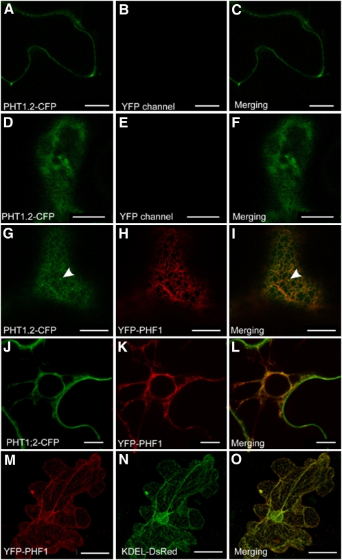
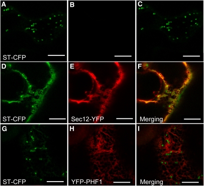
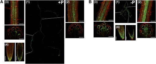
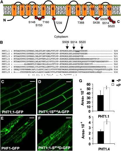
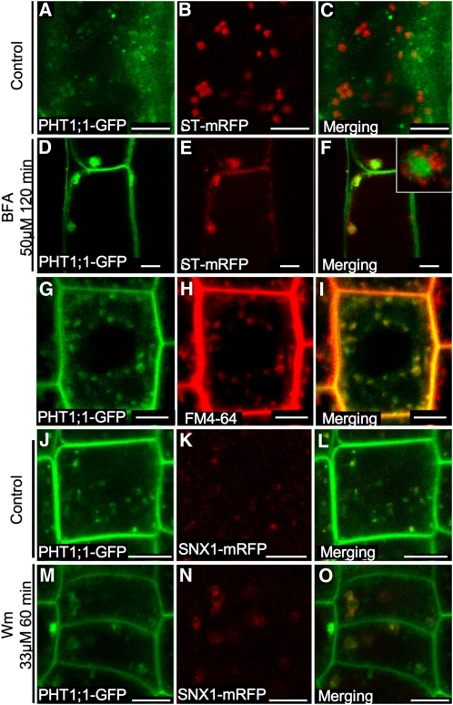
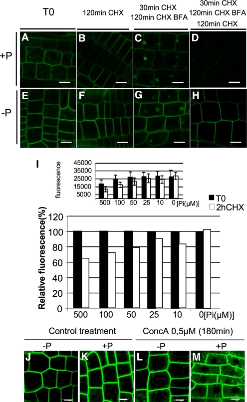
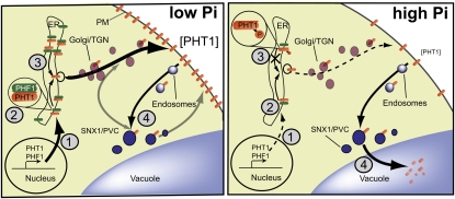
In Arabidopsis thaliana, the PHOSPHATE TRANSPORTER1 (PHT1) family encodes the high-affinity phosphate transporters. They are transcriptionally induced by phosphate starvation and require PHOSPHATE TRANSPORTER TRAFFIC FACILITATOR (PHF1) to exit the endoplasmic reticulum (ER), indicating intracellular traffic as an additional level of regulation of PHT1 activity. Our study revealed that PHF1 acts on PHT1, upstream of vesicle coat protein COPII formation, and that additional regulatory events occur during PHT1 trafficking and determine its ER exit and plasma membrane stability. Phosphoproteomic and mutagenesis analyses revealed modulation of PHT1;1 ER export by Ser-514 phosphorylation status. Confocal microscopy analysis of root tip cells showed that PHT1;1 is localized to the plasma membrane and is present in intracellular endocytic compartments. More precisely, PHT1;1 was localized to sorting endosomes associated with prevacuolar compartments. Kinetic analysis of PHT1;1 stability and targeting suggested a modulation of PHT1 internalization from the plasma membrane to the endosomes, followed by either subsequent recycling (in low Pi) or vacuolar degradation (in high Pi). For the latter condition, we identified a rapid mechanism that reduces the pool of PHT1 proteins present at the plasma membrane. This mechanism is regulated by the Pi concentration in the medium and appears to be independent of degradation mechanisms potentially regulated by the PHO2 ubiquitin conjugase. We propose a model for differential trafficking of PHT1 to the plasma membrane or vacuole as a function of phosphate concentration.


Figures

Figure 1.
Figure 1.
PHF1 Facilitates PHT1;2-CFP Targeting through the ER Compartment. Transient protein expression in N. benthamiana epidermal cells analyzed by confocal microscopy 48 h after infiltration. Transient expression of PHT1;2-CFP alone to a low (A) or high level (D) and together with YFP-PHF1 ([G] to [L]). (G) to (I) show colocalization in the ER (arrowheads indicate post-ER compartments). (J) to (L) show PHT1;2-CFP delivery to plasma membrane at cell periphery. (M) to (O) show transient coexpression of YFP-PHF1 (J) with ER marker KDEL-DsRed (K). Bars = 10 mm, except in (J) to (L), where bars = 50 μm.
Figure 2.
Figure 2.
PHF1 Is Not Associated with COPII Component Recruitment to ERES. Transient protein expression in N. benthamiana epidermal cells analyzed by confocal microscopy 48 h after infiltration. Transient expression of Golgi marker ST-CFP alone ([A] to [C]) or together with Sec12-YFP ([D] to [F]) or YFP-PHF1 ([G] to [I]). Sec12-YFP overexpression induces ST-CFP retention in ER. Bars = 10 μm.
Figure 3.
Figure 3.
PHF1 Accumulation Is Modulated by Phosphate Supply. (A) and (B) Seedlings cultivated on medium containing 500 μM (A) or 0 μM (B) Pi. (A1) and (B1) Whole root system fluorescence observed using a dissecting microscope. (A2) to (A4) and (B2) to (B4) Cell wall stained with propidium iodide (20 mg/mL) and green (GFP) fluorescence observed by confocal laser scanning microscopy. Sections are derived from Z-stack acquisitions. (A2) and (B2) Primary root. (A3) and (B3) Secondary root. (A4) and (B4) Root tip. Bars = 50 μm.
Figure 4.
Figure 4.
Targeted Mutagenesis of Putative PHT1 Phosphorylation Sites Prevents Exit from the ER. (A) Model of PHT1;1 showing potential phosphorylation sites on the cytoplasmic side of the membrane that were mutated to both Ala and Asp. Cylinders represent the 12 predicted transmembrane regions. Ser and Thr residues mutated at potential phosphorylation sites are labeled with their amino acid numbers. C, C terminus; N, N terminus. (B) Sequence alignment of the C-terminal regions of Arabidopsis PHT1 transporters. Arrows indicate the residues of PHT1;1 that were mutated (at potential phosphorylation sites). The putative ER exit site predicted for PHT1;1 is underlined. (C) to (F) Representative confocal laser scanning micrographs of the fluorescence emitted by root cells of transgenic plants expressing PHT1;1-GFP (C), PHT1;1-S514A-GFP (D), PHF1-GFP (E), and PHT1;1-S514D-GFP (F). (G) Quantitative variation of the monophosphorylated C-terminal peptides of PHT1.1 [SLEELSGEAEV(pS)HDEK] and PHT1.4 [SLEEMSGENEDNEN(pS)NNDSR] identified in proteins from root samples Pi-starved (left) or resupplied with phosphate during 2 h. Data show the mean and sd of biological triplicates with technical replicates performed. The P value corresponding to Student’s t test of significant differences between conditions were 0.0326 for PHT1.1 and 0.0006 for PHT1.4. [See online article for color version of this figure.]
Figure 5.
Figure 5.
PHT1;1 in Arabidopsis Root Tips Is Localized to Sorting Endosomes. (A) to (F) Root tip cells coexpressing PHT1;1-GFP and Golgi marker ST-mRFP in control plants ([A] to [C]) or in plants treated with 50 μM BFA for 2 h ([D] to [F]). (G) to (I) Root tip cells expressing PHT1;1-GFP were stained with the endocytic tracer FM4-64. Observations were made 15 min after incubation. (J) to (O) Root tip cells coexpressing PHT1;1-GFP and sorting endosomes marker SNX1-mRFP control ([J] to [L]) and 60 min after 33 μM Wm treatment ([M] to [O]). Bars = 5 μm.
Figure 6.
Figure 6.
Phosphate-Induced Degradation of Plasma Membrane PHT1. (A) to (M) Arabidopsis root tip cells coexpressing PHT1;1-GFP cultivated on medium containing 500 μM ([A] to [D]) or 0 μM ([E] to [H]) Pi. Images represent the following: prior to drug treatment T0 ([A] and [E]), 2 h after CHX (50 μM) treatment ([B] and [F]), 30 min CHX (50 μM) pretreatment and then 2 h CHX (50 μM) and BFA (50 μM) ([C] and [G]), and 30 min CHX (50 μM) pretreatment and then 2 h CHX (50 μM) and BFA (50 μM) followed by 2 h CHX (50 μM) washout ([D] and [H]). Bars = 10 μm. (I) Top graph shows fluorescence intensity (absolute values in arbitrary units) at T0 and after 2 h of CHX treatment of seedlings cultivated under various Pi concentrations. Bottom graph shows same data in relative level of fluorescence. For each condition, two lateral roots for six plants were observed. The fluorescence from 25 regions of interest for each sample was quantified. The mean and sd from the 300 measurements are indicated here for one of the three independent experiments performed. (J) to (M) Effect of concA treatment (0.5 μM, 3 h) on root tip cells expressing PHT1;1-GFP grown in −P and +P medium ([L] and [M]) and respective untreated control ([J] and [K]). [See online article for color version of this figure.]
Figure 7.
Figure 7.
Multiple Regulatory Events Control PHT1 Trafficking. Four different levels of control regulate PHT1 accumulation. A first one takes place at the transcriptional level (1), the second one involves PHF1 accumulation in the ER (2), the third one involves specific phosphorylation at the C terminus of PHT1 impairing PHT1 export from the ER (3), and the fourth one involves a recycling/degradation process at the level of plasma membrane (PM; 4). All these steps appear to be modulated by the Pi status of the plant. TGN, trans-Golgi network. [See online article for color version of this figure.]

Similar articles

Cited by

References

    1. Aung K., Lin S.I., Wu C.C., Huang Y.T., Su C.L., Chiou T.J. (2006). pho2, a phosphate overaccumulator, is caused by a nonsense mutation in a microRNA399 target gene. Plant Physiol. 141: 1000–1011 - PMC - PubMed
    1. Barber S.A., Walker J.M., Vasey E.H. (1963). Mechanisms for movement of plant nutrients from soil and fertilizer to plant root. J. Agric. Food Chem. 11: 204–207
    1. Bari R., Datt Pant B., Stitt M., Scheible W.R. (2006). PHO2, microRNA399, and PHR1 define a phosphate-signaling pathway in plants. Plant Physiol. 141: 988–999 - PMC - PubMed
    1. Bayle V., Nussaume L., Bhat R.A. (2008). Combination of novel GFP mutant TSapphire and DsRed variant mOrange to set up a versatile in planta FRET-FLIM assay. Plant Physiol. 148: 51–60 - PMC - PubMed
    1. Bolte S., Cordelières F.P. (2006). A guided tour into subcellular colocalization analysis in light microscopy. J. Microsc. 224: 213–232 - PubMed

Publication types

MeSH terms