. 2011 Jun;9(6):746-56.
doi: 10.1158/1541-7786.MCR-11-0112. Epub 2011 Apr 26.

Pharmacologic inhibition of ALK5 causes selective induction of terminal differentiation in mouse keratinocytes expressing oncogenic HRAS

Affiliations

Pharmacologic inhibition of ALK5 causes selective induction of terminal differentiation in mouse keratinocytes expressing oncogenic HRAS

Lauren Mordasky Markell et al. Mol Cancer Res. 2011 Jun.

Abstract

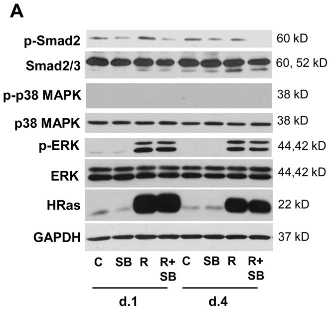
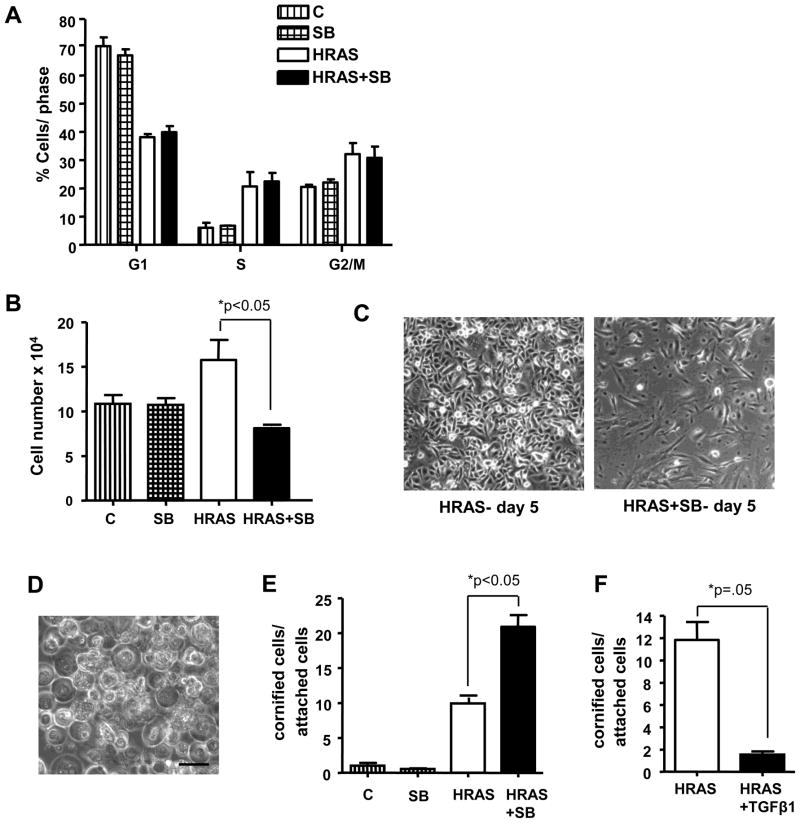
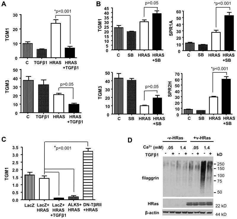
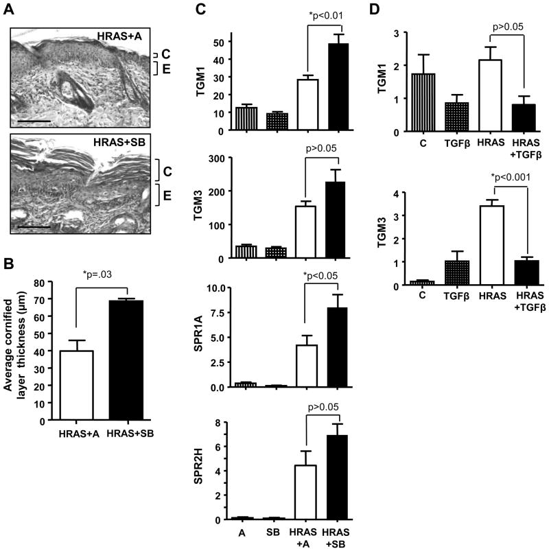
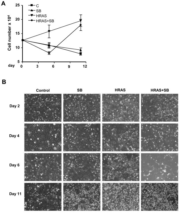
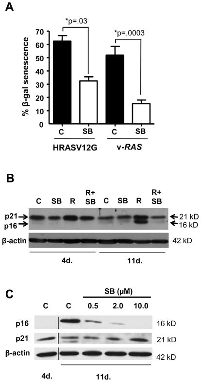
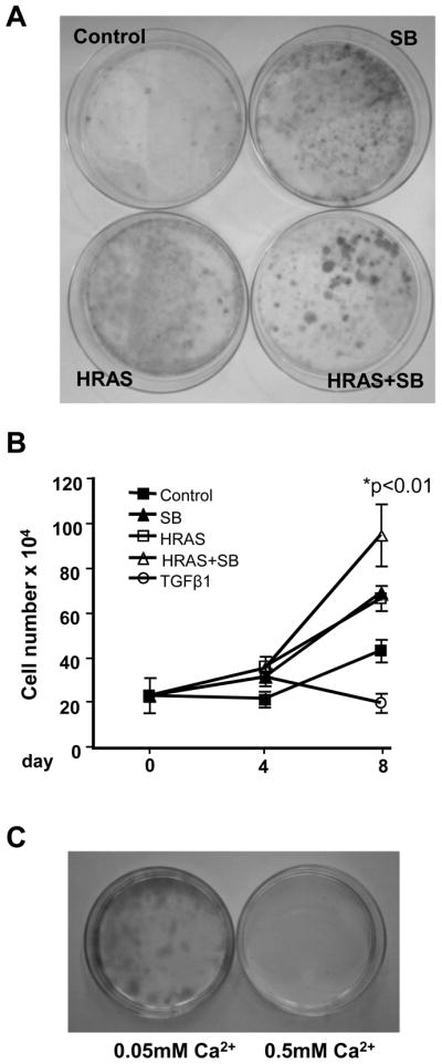
TGFβ has both tumor suppressive and oncogenic roles in cancer development. We previously showed that SB431542 (SB), a small molecule inhibitor of the TGFβ type I receptor (ALK5) kinase, suppressed benign epidermal tumor formation but enhanced malignant conversion. Here, we show that SB treatment of primary K5rTA/tetORASV12G bitransgenic keratinocytes did not alter HRASV12G-induced keratinocyte hyperproliferation. However, continuous SB treatment significantly enhanced HRASV12G-induced cornified envelope formation and cell death linked to increased expression of enzymes transglutaminase (TGM) 1 and TGM3 and constituents of the cornified envelope small proline-rich protein (SPR) 1A and SPR2H. In contrast, TGFβ1 suppressed cornified envelope formation in HRASV12G keratinocytes. Similar results were obtained in HRASV12G transgenic mice treated topically with SB or by coexpressing TGFβ1 and HRASV12G in the epidermis. Despite significant cell death, SB-resistant HRASV12G keratinocytes repopulated the primary culture that had overcome HRas-induced senescence. These cells expressed reduced levels of p16(ink4a) and were growth stimulated by SB but remained sensitive to a calcium-induced growth arrest. Together these results suggest that differential responsiveness to cornification may represent a mechanism by which pharmacologic blockade of TGFβ signaling can inhibit the outgrowth of preneoplastic lesions but may cause a more progressed phenotype in a separate keratinocyte population.


Conflict of interest statement

Conflict of Interest: The authors declare no conflict of interest.

Figures

Figure 1
Figure 1
SB431542 inhibits Smad2 phosphorylation in bitransgenic keratinocytes. A, Immunoblot showing induction of HRASV12G and Erk1/2 phosphorylation with Dox and reduction in p-Smad2 levels in HRASV12G-expressing and control keratinocytes treated with SB for indicated times. SB does not inhibit phosphorylation of ERK and no increase in p38 MAPK phosphorylation was observed with HRASV12G induction. Protein isolation and analysis as described in Materials and Methods, with GAPDH used as a loading control. N=2. C, Control; R, HRASV12G expression induced by Dox.
Figure 2
Figure 2
SB431542 enhances terminal differentiation in HRASV12G-expressing keratinocytes but does not affect cell proliferation. A, 2-color flow cytometric analysis of cell proliferation after 4 days of SB treatment shows no significant effect on cell cycle of either control or HRASV12G keratinocytes. Cells were stained with BrdU/PI and analyzed according to Materials and Methods. n=2, N=2. B, Attached cell number of HRASV12G keratinocytes is significantly reduced after 5 days of continuous treatment with SB. n=3, N=2. C, Photomicrographs showing effect of SB on bitransgenic cell monolayers after 5 days. HRASV12G keratinocytes alone (left) and HRASV12G + SB (right) on day 5. 100x magnification. D, Photomicrograph of floating cornified cells from SB treated HRASV12G keratinocytes. 200x magnification, scale bar represents 50μm. E, Enhanced formation of cornified envelopes in keratinocytes expressing HRASV12G treated with SB. Floating cells counted in triplicate by cornified envelope assay and expressed relative to attached cells on day 5. n=3, N=3. F, TGFβ1 blocks cornified envelope formation in HRASV12G- expressing keratinocytes. HRASV12G was induced for 3 days followed by an additional 2 days in the presence or absence of 1ng/ml TGFβ1. Cornified cells were measured as described in Materials in Methods and expressed relative to total attached cells. All keratinocytes were cultured and treated in 0.05mM Ca2+ media. n=3, N=2. C, Control; HRAS, HRASV12G expression induced by Dox. Values with * are significantly different at p <0.05. Error bars represent standard error of the mean.
Figure 3
Figure 3
TGFβ signaling regulates expression of terminal differentiation genes in preneoplastic keratinocytes. A, Reduced expression of TGM1 and TGM3 in HRASV12G keratinocytes treated with TGFβ1. Bitransgenic keratinocytes were treated with Dox for 24h and then a further 12h with Dox ±TGFβ1 prior to isolation of RNA. n=4, N=2. B, SB increases expression of terminal differentiation genes in HRASV12G keratinocytes. Bitransgenic keratinocytes were treated for 4 days with Dox±SB prior to isolation of RNA. n=4, N=3. C, Infection of HRASV12G-expressing keratinocytes with adenoviruses expressing either a constitutively activated TGFβ type I receptor (ALK5) or a dominant negative TGFβ type II receptor inhibits and induces TGM1 expression, respectively. RNA was isolated from cells 24h after adenoviral infection. n=3, N=2. D, TGFβ1 blocks elevated Ca2+ induced expression of filaggrin in v-HRas infected keratinocytes. Protein extracts were isolated 24h after switching v-HRas infected keratinocytes to 0.5mM Ca2+ in the presence or absence of 1ng/ml TGFβ1, and immunoblotted for the indicated proteins. β-actin was used as a loading control. N=1. C, control; HRAS, HRASV12G expression induced by Dox. Values with * are significantly different at p <0.05. Error bars represent standard error of the mean.
Figure 4
Figure 4
TGFβ signaling pathway regulates expression of terminal differentiation markers in vivo. A, Photomicrographs showing increased thickness of the cornified layer in bitransgenic Involucrin tTA/tetORASV12G mice treated with SB (10.0μM in 200μl acetone) for 5 days. Mice were removed from Dox to induce HRASV12G expression. Magnification 200x, scale bar represents 50 μm. n=5. C, Cornified layer; E, Epidermis. B, Quantification of cornified layer thickness, measured at magnification 200x, 30 measurements/slide. n=5. C, Topical SB (10.0μM) treatment of InvtTA/tetORASV12G mice significantly increases TGM1 and SPR1A gene expression, with similar trends for TGM3 and SPR2H. n=5. D, Co-induction of TGFβ1 with HRASV12G in triple transgenic mice reduces expression of TGM1 and TGM3 compared to HRASV12G alone. n=5. A, Acetone; HRAS, HRASV12G expression induced by Dox removal; TGFβ, tetOTGFβ. Values with * are significantly different at p <0.05. Error bars represent standard error of the mean.
Figure 5
Figure 5
Continued proliferation of HRASV12G keratinocytes resistant to SB431542-induced cornification. A, Growth curve showing expansion of SB resistant cells and recovery of attached cell numbers after 11 days continuous treatment. n=3, N=2. B, Photomicrographs of bitransgenic monolayers cultured for 11 days with and without HRASV12G expression and SB, showing loss of attached cells at day 6, re-growth by day 11 and lack of effect of SB on keratinocytes not expressing oncogenic HRas. C, Control, bitransgenic keratinocytes without Dox or SB; HRAS, HRASV12G expression induced by Dox.
Figure 6
Figure 6
Reduced Senescence in HRas-expressing keratinocytes treated long term with SB431542. A, HRASV12G and v-HRas expressing Balb/c keratinocytes treated for 11 days with SB have reduced percentage of senescent cells compared to untreated HRas expressing controls. n=3, N=2. B, Senescence-associated increase in p16ink4a is blocked in HRASV12G keratinocytes cultured in SB. Blot was probed for p21waf1 followed by p16ink4a without stripping. N=2. C, Dose dependent inhibition of p16ink4a protein expression by SB in v-HRas infected keratinocytes. Retrovirally infected Balb/c keratinocytes were treated with the indicated dose of SB continuously for 11 days prior to isolation of cell extract for immunoblotting. No change in p15ink4b was observed (not shown). β-actin was used as a loading control. N=2. C, Control; R, HRASV12G expression induced by Dox. Values with * are significantly different at p <0.05. Error bars represent standard error of the mean.
Figure 7
Figure 7
SB431542 resistant cells arise from a subpopulation of HRASV12G keratinocytes stimulated to proliferate by SB. A, HRASV12G keratinocytes plated at low density and treated with SB (2.0 μM) for 11 days form large colonies while cells expressing HRASV12G or treated with SB alone do not. N=3. B, Passaged resistant cells are growth stimulated by re-expression of HRASV12G and SB. A mass culture of SB resistant cells was passaged twice before plating in media without supplements for 2 days followed by treatment as indicated, and attached cell number was determined at the indicated days. There was a significant increase in cell number in cells expressing HRASV12G+SB compared to HRASV12G expression alone. n=3. N=2. C, Passaged HRASV12G+SB resistant keratinocytes can grow clonally in proliferation media (0.05 mM Ca2+) with continued Dox and SB treatment (left) but are unable to form colonies in differentiation media (0.5mM Ca2+ media, right). No colonies formed in either growth condition when cells were cultured with Dox or SB alone or without any treatment. Passaged cells were seeded at clonal density in the presence of Dox and SB to allow attachment and then switched to the indicated conditions. N=4. HRAS, HRASV12G expression induced by Dox; TGFβ1, 1ng/ml. Values with * are significantly different at p <0.05. Error bars represent standard error of the mean.

References

    1. Fowlis DJ, Cui W, Johnson SA, Balmain A, Akhurst RJ. Altered epidermal cell growth control in vivo by inducible expression of transforming growth factor β1 in the skin of transgenic mice. Cell Growth Differ. 1996;7:679–87. - PubMed
    1. Blessing M, Nanney LB, King LE, Hogan BL. Chemical skin carcinogenesis is prevented in mice by the induced expression of a TGF-β related transgene. Teratog Carcinog Mutagen. 1995;15:11–21. - PubMed
    1. Honjo Y, Bian Y, Kawakami K, Molinolo A, Longenecker G, Boppana R, et al. TGF-beta receptor I conditional knockout mice develop spontaneous squamous cell carcinoma. Cell Cycle. 2007;6:1360–6. - PubMed
    1. Bian Y, Terse A, Du J, Hall B, Molinolo A, Zhang P, et al. Progressive tumor formation in mice with conditional deletion of TGF-beta signaling in head and neck epithelia is associated with activation of the PI3K/Akt pathway. Cancer Res. 2009;69:5918–26. - PMC - PubMed
    1. Amendt C, Schirmacher P, Weber H, Blessing M. Expression of a dominant negative type II TGF-β receptor in mouse skin results in an increase in carcinoma incidence and an acceleration of carcinoma development. Oncogene. 1998;17:25–34. - PubMed

Publication types

MeSH terms

Substances