. 2011 May 10;108(19):7832-7.
doi: 10.1073/pnas.1101379108. Epub 2011 Apr 26.

Selective activation of the transcription factor ATF6 mediates endoplasmic reticulum proliferation triggered by a membrane protein

Affiliations

Selective activation of the transcription factor ATF6 mediates endoplasmic reticulum proliferation triggered by a membrane protein

Jessica Maiuolo et al. Proc Natl Acad Sci U S A. .

Abstract

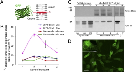
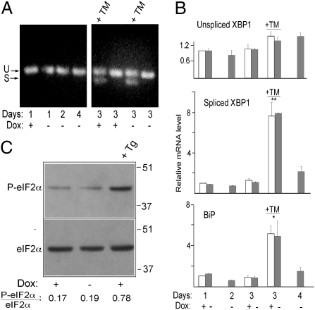
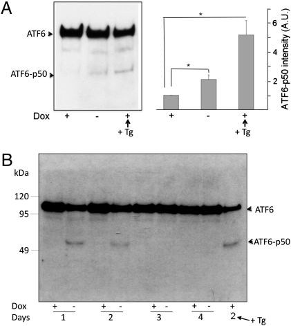
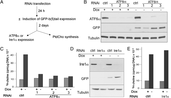
It is well known that the endoplasmic reticulum (ER) is capable of expanding its surface area in response both to cargo load and to increased expression of resident membrane proteins. Although the response to increased cargo load, known as the unfolded protein response (UPR), is well characterized, the mechanism of the response to membrane protein load has been unclear. As a model system to investigate this phenomenon, we have used a HeLa-TetOff cell line inducibly expressing a tail-anchored construct consisting of an N-terminal cytosolic GFP moiety anchored to the ER membrane by the tail of cytochrome b5 [GFP-b(5)tail]. After removal of doxycycline, GFP-b(5)tail is expressed at moderate levels (1-2% of total ER protein) that, nevertheless, induce ER proliferation, as assessed both by EM and by a three- to fourfold increase in phosphatidylcholine synthesis. We investigated possible participation of each of the three arms of the UPR and found that only the activating transcription factor 6 (ATF6) arm was selectively activated after induction of GFP-b(5)tail expression; peak ATF6α activation preceded the increase in phosphatidylcholine synthesis. Surprisingly, up-regulation of known ATF6 target genes was not observed under these conditions. Silencing of ATF6α abolished the ER proliferation response, whereas knockdown of Ire1 was without effect. Because GFP-b(5)tail lacks a luminal domain, the response we observe is unlikely to originate from the ER lumen. Instead, we propose that a sensing mechanism operates within the lipid bilayer to trigger the selective activation of ATF6.


Conflict of interest statement

The authors declare no conflict of interest.

Figures

Fig. 1.
Fig. 1.
Moderate levels of GFP-b(5)tail induce PtdCho synthesis. (A) Schematic representation of GFP-b(5)tail [a detailed description of the construct is presented elsewhere (15)]. (B) PtdCho synthesis in induced or noninduced cells. GFP-b(5)tail–transfected and nontransfected HeLa-TetOff cells, grown in the presence or absence of Dox, were incubated with 3H-choline on the indicated days. (C) Evaluation of GFP-b(5)tail expression induced by Dox removal. (Lower) Immunoblot of increasing amounts of standard protein (lanes 1–3) and of 40 μg of total protein from cells grown in the absence of Dox for the indicated times (lanes 4–6). A heavily overexposed blot of the 4-d sample in comparison with cells grown in the presence of Dox is shown in lanes 7 and 8. (Upper) Portion of the two blots stained with Amido Black to check for protein load. (D) Confocal analysis of HeLa-TetOff cells expressing GFP-b(5)tail after 3 d of exposure to Dox-free medium. (Inset) Cell indicated with the asterisk at higher magnification. (Scale bars: 20 μm; Inset, 5 μm.)
Fig. 2.
Fig. 2.
Absence of ROS generation or Ca2+ leakage from the ER in GFP-b(5)tail–expressing cells. (A) Determination of ROS production in cells grown with or without Dox for the indicated days. H2O2 was added to one sample immediately before carrying out the fluorimeter readings. Shown are the 30-min readings corrected for the values recorded at the beginning of the registration, according to the equation [(F30 − F0)/F0] × 100. (B and C) Intracellular [Ca2+] measurements on induced and noninduced cells. (B) Representative 340:380 traces of single cells (each shown in a different color) from the two cultures, under basal conditions (first few seconds of each trace), during exposure to Tg and after addition of EGTA. (C) Average 340:380 values ± SD under basal conditions (Left) and peak amplitude of response to Tg (Right) for the two cell populations. The number of cells analyzed for each condition is indicated within the columns. A.U., arbitrary units.
Fig. 3.
Fig. 3.
Absence of XBP1 splicing or PERK activation in GFP-b(5)tail–expressing cells. (A) Evaluation of XBP1 mRNA splicing by RT-PCR on cells maintained in the presence or absence of Dox for the indicated times. The +TM lanes contained reactions from cells treated for 4 h with the drug (6 μg/mL). U and S indicate the products of unspliced and spliced XBP1 mRNA, respectively. (B) Quantification of XBP1 and BiP mRNAs by RT-qPCR. Values are normalized to those of the +Dox cells at 1 d after replating. Shown are mean values ± SEM from three independent experiments. Statistical probability, indicated by the asterisks, refers to TM-treated cells vs. all untreated cells (*P < 0.05; **P < 0.01 by one-way ANOVA followed by Tukey's posttest). (C) Total and P-eIF2α detected by immunoblotting. Lysates were from GFP-b(5)tail–HeLa-TetOff cells grown in the presence or absence of Dox for 48 h. Cells were treated with 300 nM Tg for 30 min where indicated. The ratio of P-eIF2α to total eIF2α is indicated below the lanes.
Fig. 4.
Fig. 4.
GFP-b(5)tail expression activates ATF6α. (A) Immunoblotting analysis of FLAG-tagged ATF6α. GFP-b(5)tail–HeLa-TetOff cells were transiently transfected with a plasmid coding for FLAG-tagged ATF6α and maintained in culture for 48 h in the presence or absence of Dox. (Left) Representative blot. (Right) Densitometric analysis of blots from three independent experiments. Mean values ± SEM are shown (*P = 0.04 and *P = 0.02 for −Dox and for +Dox + Tg cells, respectively, vs. +Dox cells by Student's two-tailed t test after logarithmic transformation of the densitometric readings). A.U., arbitrary units. (B) Time course of endogenous ATF6α cleavage in GFP-b(5)tail–HeLa-TetOff cells assessed by immunoblotting. In A and B, cells were treated with 300 nM Tg for 1 h where indicated.
Fig. 5.
Fig. 5.
Silencing of ATF6α but not of Ire1α deletes the increase in PtdCho synthesis driven by GFP-b(5)tail. (A) Design of the experiments. (B) Silencing of ATF6α by three different siRNAs in induced and noninduced cells, shown by immunoblotting. (C) Stimulation of PtdCho synthesis by GFP-b(5)tail expression is strongly reduced after transfection with the three different ATF6α siRNAs. (D) Immunoblotting shows silencing of Ire1α by a pool of three siRNAs, both in induced and noninduced cells. (E) PtdCho synthesis stimulated by GFP-b(5)tail expression is unaffected by transfection with the Ire1α siRNA pool. ctrl, control.

References

    1. Ron D, Walter P. Signal integration in the endoplasmic reticulum unfolded protein response. Nat Rev Mol Cell Biol. 2007;8:519–529. - PubMed
    1. Cox JS, Chapman RE, Walter P. The unfolded protein response coordinates the production of endoplasmic reticulum protein and endoplasmic reticulum membrane. Mol Biol Cell. 1997;8:1805–1814. - PMC - PubMed
    1. Sriburi R, Jackowski S, Mori K, Brewer JW. XBP1: A link between the unfolded protein response, lipid biosynthesis, and biogenesis of the endoplasmic reticulum. J Cell Biol. 2004;167:35–41. - PMC - PubMed
    1. Bommiasamy H, et al. ATF6alpha induces XBP1-independent expansion of the endoplasmic reticulum. J Cell Sci. 2009;122:1626–1636. - PMC - PubMed
    1. Orrenius S, Ericsson JL, Ernster L. Phenobarbital-induced synthesis of the microsomal drug-metabolizing enzyme system and its relationship to the proliferation of endoplasmic membranes. A morphological and biochemical study. J Cell Biol. 1965;25:627–639. - PMC - PubMed

Publication types

MeSH terms

Substances