

Dedifferentiation of immortalized human podocytes in response to transforming growth factor-β: a model for diabetic podocytopathy
- PMID: 21521871
- PMCID: PMC3114395
- DOI: 10.2337/db10-1110
Dedifferentiation of immortalized human podocytes in response to transforming growth factor-β: a model for diabetic podocytopathy
Abstract
Objective: Diabetic nephropathy is associated with dedifferentiation of podocytes, losing the specialized features required for efficient glomerular function and acquiring a number of profibrotic, proinflammatory, and proliferative features. These result from tight junction and cytoskeletal rearrangement, augmented proliferation, and apoptosis.
Research design and methods: Experiments were performed in conditionally immortalized human podocytes developed by transfection with the temperature-sensitive SV40-T gene. Cells were then cultured in the presence of transforming growth factor (TGF)-β1 or angiotensin II in the presence or absence of a selective inhibitor of the TGF-β type I receptor kinase, SB-431542. Gene and protein expression were then examined by real-time RT-PCR and immunofluorescence, and correlated with changes observed in vivo in experimental diabetes.
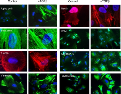
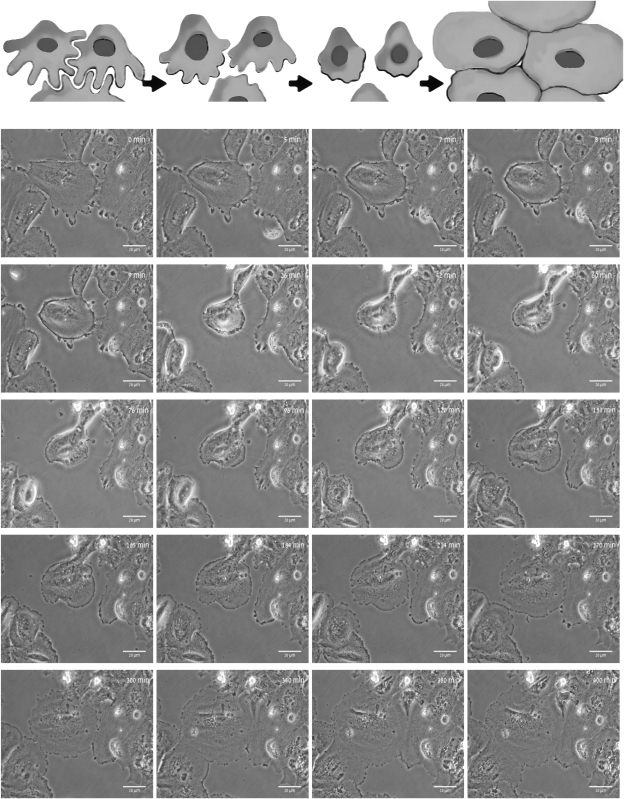
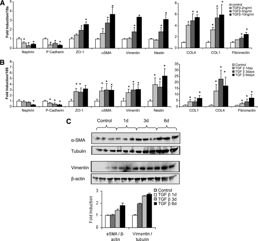
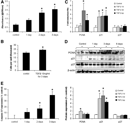
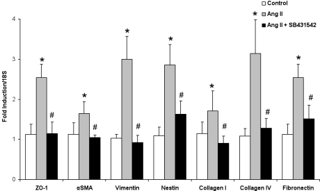
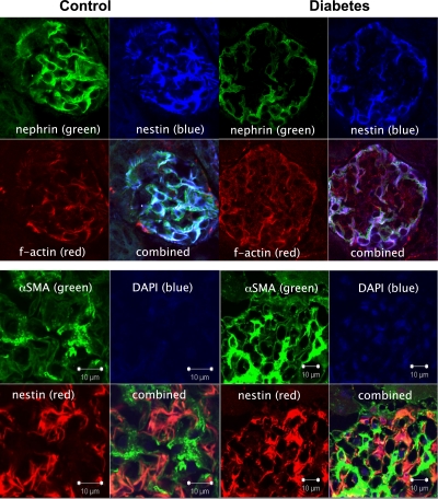
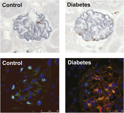
Results: Treatment of cells with TGF-β1 resulted in dynamic changes in their morphology, starting with retraction and shortening of foot processes and finishing with the formation of broad and complex tight junctions between adjacent podocytes. This dedifferentiation was also associated with dose- and time-dependent reduction in the expression of glomerular epithelial markers (nephrin, p-cadherin, zonnula occludens-1) and increased expression of mesenchymal markers (α-smooth muscle actin, vimentin, nestin), matrix components (fibronectin, collagen I, and collagen IV α3), cellular proliferation, and apoptosis. The induction of diabetes in mice was also associated with similar changes in morphology, protein expression, and proliferation in glomerular podocytes.
Conclusions: In response to TGF-β and other TGF-dependent stimuli, mature podocytes undergo dedifferentiation that leads to effacement of foot processes, morphologic flattening, and increased formation of intercellular tight junctions. This simplification of their phenotype to a more embryonic form is also associated with reentry of mature podocytes into the cell cycle, which results in enhanced proliferation and apoptosis. These "pathoadaptive" changes are seen early in the diabetic glomerulus and ultimately contribute to albuminuria, glomerulosclerosis, and podocytopenia.
Figures

References
-
- Ziyadeh FN, Wolf G. Pathogenesis of the podocytopathy and proteinuria in diabetic glomerulopathy. Curr Diabetes Rev 2008;4:39–45 - PubMed
-
- Wolf G, Chen S, Ziyadeh FN. From the periphery of the glomerular capillary wall toward the center of disease: podocyte injury comes of age in diabetic nephropathy. Diabetes 2005;54:1626–1634 - PubMed
-
- Li JJ, Kwak SJ, Jung DS, et al. Podocyte biology in diabetic nephropathy. Kidney Int Suppl 2007:S36–S42 - PubMed
-
- Salmon AH, Toma I, Sipos A, et al. Evidence for restriction of fluid and solute movement across the glomerular capillary wall by the subpodocyte space. Am J Physiol Renal Physiol 2007;293:F1777–F1786 - PubMed
Publication types
MeSH terms
Substances
LinkOut - more resources
Full Text Sources
Other Literature Sources
Medical
Molecular Biology Databases
