. 2011 Sep;19(9):965-73.
doi: 10.1038/ejhg.2011.63. Epub 2011 Apr 27.

The DFNA5 gene, responsible for hearing loss and involved in cancer, encodes a novel apoptosis-inducing protein

Affiliations

The DFNA5 gene, responsible for hearing loss and involved in cancer, encodes a novel apoptosis-inducing protein

Ken Op de Beeck et al. Eur J Hum Genet. 2011 Sep.

Abstract

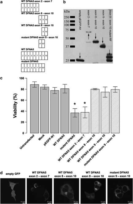
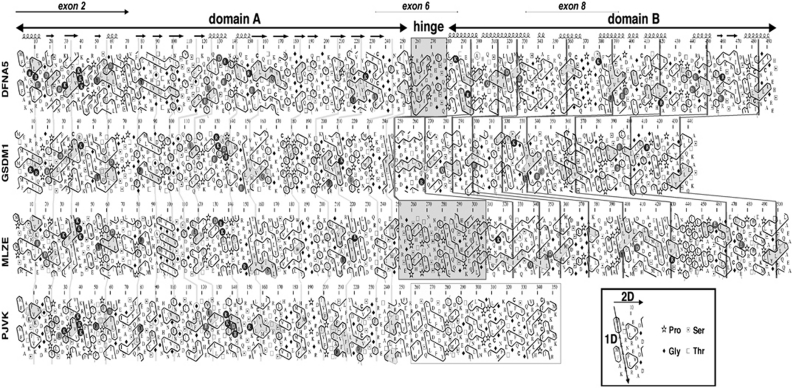
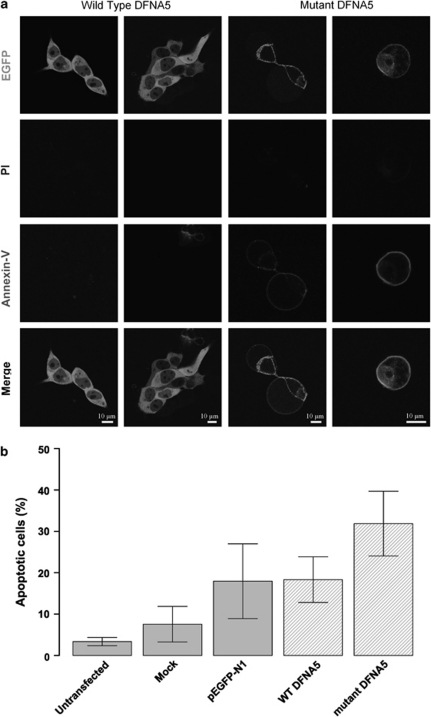
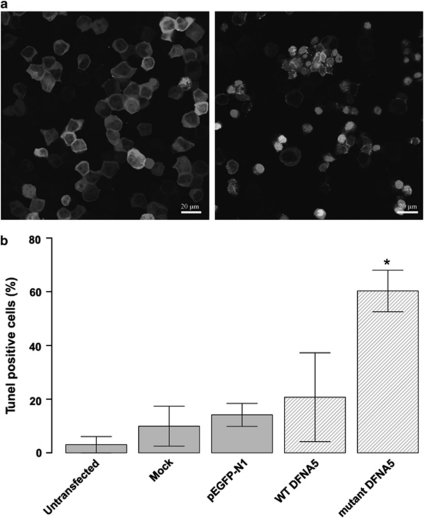
DFNA5 was first identified as a gene causing autosomal dominant hearing loss (HL). Different mutations have been found, all exerting a highly specific gain-of-function effect, in which skipping of exon 8 causes the HL. Later reports revealed the involvement of the gene in different types of cancer. Epigenetic silencing of DFNA5 in a large percentage of gastric, colorectal and breast tumors and p53-dependent transcriptional activity have been reported, concluding that DFNA5 acts as a tumor suppressor gene in different frequent types of cancer. Despite these data, the molecular function of DFNA5 has not been investigated properly. Previous transfection studies with mutant DFNA5 in yeast and in mammalian cells showed a toxic effect of the mutant protein, which was not seen after transfection of the wild-type protein. Here, we demonstrate that DFNA5 is composed of two domains, separated by a hinge region. The first region induces apoptosis when transfected in HEK293T cells, the second region masks and probably regulates this apoptosis inducing capability. Moreover, the involvement of DFNA5 in apoptosis-related pathways in a physiological setting was demonstrated through gene expression microarray analysis using Dfna5 knockout mice. In view of its important role in carcinogenesis, this finding is expected to lead to new insights on the role of apoptosis in many types of cancer. In addition, it provides a new line of evidence supporting an important role for apoptosis in monogenic and complex forms of HL.


Figures

Figure 1
Figure 1
(a) Graphical representation of the insert content in relation to the construct's name. An exon is indicated by a rectangle containing the relevant exon number. The alternative reading frame used in the aberrant tail of mutant DFNA5 is indicated with an asterisk. (b) Western blot analysis of selected DFNA5 constructs demonstrating that all constructs are expressed when transfected in HEK293T cells (using a pEGFP-N1 vector). Arrows point to the relevant position of the fusion proteins. Some degradation products are visible in lanes 2, 3 and 5. (c) Cell viability measurements 21 h post-transfection. Graphical bars represent the percentage of viable cells. An asterisk denotes statistically significant differences (P<0.05) between transfections with the construct of interest and the WT construct. Control measurements are depicted in light gray colored bars, while constructs specific for this experiment are indicated with hatched bars. This series of transfections demonstrates that the toxic part of DFNA5 is located in the first part of DFNA5. Interestingly, this indicates that the motif that is responsible for toxicity is shared by WT and mutant DFNA5. (d) Subcellular localization of EGFP fusion proteins in HEK293T cells. Transfection of empty pEGFP-N1 vector results in an evenly distributed expression pattern throughout the cytosol. The first part of DFNA5 (exon 2–exon 7) is localized mainly at the plasma membrane. Cell blebbing is seen in cells transfected with this construct. A similar pattern was observed when mutant DFNA5 was transfected in HEK293T cells. Both WT DFNA5 exon 8–exon 10 and WT DFNA5 exon 9–exon 10 are localized in the cytoplasm, near the nucleus. Cells transfected with the mutant DFNA5 exon 9–exon 10 construct show expression in the endoplasmic reticulum (see also Supplementary Figure 1). A full color version of this figure can be found in the html version of this paper.
Figure 2
Figure 2
HCA analysis of DFNA5, GSDM1, MLZE and PJVK. The sequences are shown on a duplicated α-helical net, in which hydrophobic residues (VILFMYW) are contoured, forming clusters, which mainly correspond to the internal faces of regular secondary structures. The way to read the sequences (1D) and secondary structures (2D) as well as special symbols are indicated in the inset. Conserved hydrophobic residues are shaded in gray, the vertical lines indicating the correspondences between the different sequences. Noticeable identities/similarities outside the hydrophobic clusters are encircled in gray. The predicted globular domains (A and B) are depicted with arrows. A hinge of variable length (gray box) separates domain A from domain B. Predicted secondary structures of these globular domains are indicated at the top. Domain B does not exist in PJVK, in which it is replaced by a shorter domain (contoured in gray), likely containing a zinc-finger domain. A full color version of this figure can be found in the html version of this paper.
Figure 3
Figure 3
(a) Confocal images of annexin V-Cy5-stained cell transfections. Cells transfected with WT DFNA5 are negative for PI and annexin V-Cy5. Cells transfected with mutant DFNA5 are negative for PI, but show clear annexin V at the plasma membrane, which is suggestive for apoptotic cell death. All cells were harvested 16 h post-transfection. A full color version of this figure can be found in the html version of this paper. (b) Flow cytometric quantification of annexin V-Cy5-stained transfected cells. Cells were harvested 16 h post-transfection. Percentages of apoptotic cells within the total cell population are shown. The total apoptotic fraction was calculated as the sum of the early (annexin V+ PI−) and late (annexin V+ PI+) apoptotic fractions.
Figure 4
Figure 4
(a) Confocal images of WT DFNA5 (left pane of image)- versus mutant DFNA5 (right pane of image)-transfected HEK293T cells, stained with the in situ cell death detection kit, TM Red. TUNEL-positive cells show bright staining of the nucleus. A full color version of this figure can be found in the html version of this paper. (b) Quantitative measurement of TUNEL-positive cells relative to the total cell population. Cells transfected with mutant DFNA5 show significantly more apoptosis compared to controls. All cells were harvested 16 h post-transfection.

References

    1. Van Laer L, Huizing EH, Verstreken M, et al. Nonsyndromic hearing impairment is associated with a mutation in DFNA5. Nat Genet. 1998;20:194–197. - PubMed
    1. Park HJ, Cho HJ, Baek JI, et al. Evidence for a founder mutation causing DFNA5 hearing loss in East Asians. J Hum Genet. 2009;55:59–62. - PMC - PubMed
    1. Cheng J, Han DY, Dai P, et al. A novel DFNA5 mutation, IVS8+4 A>G, in the splice donor site of intron 8 causes late-onset non-syndromic hearing loss in a Chinese family. Clin Genet. 2007;72:471–477. - PubMed
    1. Bischoff AMLC, Luijendijk MWJ, Huygen PLM, et al. A second mutation identified in the DFNA5 gene in a Dutch family. A clinical and genetic evaluation. Audiol Neuro-otol. 2004;9:34–36. - PubMed
    1. Yu C, Meng X, Zhang S, Zhao G, Hu L, Kong X. A 3-nucleotide deletion in the polypyrimidine tract of intron 7 of the DFNA5 gene causes nonsyndromic hearing impairment in a Chinese family. Genomics. 2003;82:575–579. - PubMed

Publication types

Substances