

Bacterial diversity in persistent periapical lesions on root-filled teeth
- PMID: 21523213
- PMCID: PMC3077005
- DOI: 10.3402/jom.v1i0.1946
Bacterial diversity in persistent periapical lesions on root-filled teeth
Abstract
Background: The purpose of this study was to analyze the bacterial diversity in persistent apical lesions on root-filled teeth by using culture-independent molecular methods.
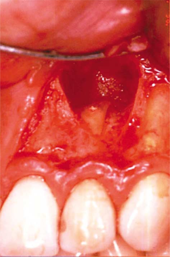
Design: Twenty surgically removed apical lesions from therapy-resistant teeth were examined for the presence of bacterial DNA using PCR targeting the 16s ribosomal RNA gene, followed by cloning and sequencing.
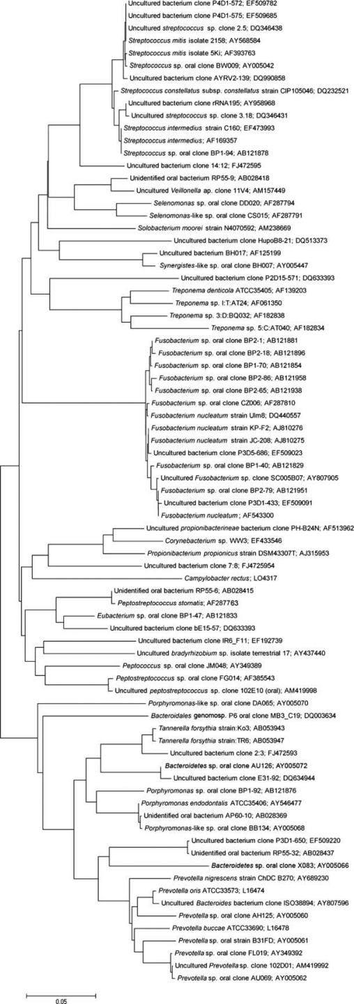
Results: Bacterial DNA was detected in 17 of the 20 samples (85%). A total of 236 clones were analyzed. Seven different bacterial phyla were represented and a total of 75 different bacterial taxa were identified; 36% of the species have not yet been cultivated. Commonly detected bacterial species included Fusobacterium spp., Prevotella spp., Tannerella forsythia, Porphyromonas endodontalis, Treponema denticola, Bacteroidetes spp., Peptostreptococcus spp., and Streptococcus spp.
Conclusions: A wide range of bacteria was identified in periapical lesions on therapy-resistant teeth. These bacteria may contribute in the etiology of periapical infection and impede healing of these lesions.
Keywords: 16s ribosomal RNA; bacterial phyla; endodontic infection; sequencing; therapy-resistant teeth.
Figures
Similar articles
-
Microbial analysis of canals of root-filled teeth with periapical lesions using polymerase chain reaction.J Endod. 2008 May;34(5):537-40. doi: 10.1016/j.joen.2008.01.016. Epub 2008 Mar 4. J Endod. 2008. PMID: 18436030
-
Microbiological examination of infected dental root canals.Oral Microbiol Immunol. 2004 Apr;19(2):71-6. doi: 10.1046/j.0902-0055.2003.00116.x. Oral Microbiol Immunol. 2004. PMID: 14871344
-
Evaluation of Bacteriological Profile in the Apical Root Segment of the Patients with Primary Apical Periodontitis.J Contemp Dent Pract. 2017 Jan 1;18(1):44-48. doi: 10.5005/jp-journals-10024-1986. J Contemp Dent Pract. 2017. PMID: 28050984
-
Microbial community in persistent apical periodontitis: a 16S rRNA gene clone library analysis.Int Endod J. 2015 Aug;48(8):717-28. doi: 10.1111/iej.12361. Epub 2014 Sep 16. Int Endod J. 2015. PMID: 25088120
-
Exploring bacterial diversity of endodontic microbiota by cloning and sequencing 16S rRNA.J Endod. 2011 Jul;37(7):922-6. doi: 10.1016/j.joen.2011.04.007. J Endod. 2011. PMID: 21689545
Cited by
-
As-yet-uncultivated oral bacteria: breadth and association with oral and extra-oral diseases.J Oral Microbiol. 2013 May 23;5. doi: 10.3402/jom.v5i0.21077. Print 2013. J Oral Microbiol. 2013. PMID: 23717756 Free PMC article.
-
Periapical bacterial disinfection is critical for dental pulp regenerative cell therapy in apical periodontitis in dogs.Stem Cell Res Ther. 2024 Jan 17;15(1):17. doi: 10.1186/s13287-023-03628-6. Stem Cell Res Ther. 2024. PMID: 38229184 Free PMC article.
-
Factors that cause endodontic failures in general practices in Japan.BMC Oral Health. 2018 Apr 27;18(1):70. doi: 10.1186/s12903-018-0530-6. BMC Oral Health. 2018. PMID: 29703201 Free PMC article.
-
Prevalence of unculturable bacteria in the periapical abscess: A systematic review and meta-analysis.PLoS One. 2021 Aug 5;16(8):e0255485. doi: 10.1371/journal.pone.0255485. eCollection 2021. PLoS One. 2021. PMID: 34351963 Free PMC article.
-
Pyrosequencing as a tool for better understanding of human microbiomes.J Oral Microbiol. 2012;4. doi: 10.3402/jom.v4i0.10743. Epub 2012 Jan 23. J Oral Microbiol. 2012. PMID: 22279602 Free PMC article.
References
-
- Eriksen HM, Kirkevang LL, Petersson K. Endodontic epidemiology and treatment outcome: general considerations. Endod Topics. 2002;2:1–9.
-
- Bergström J, Eliasson S, Ahlberg KF. Periapical status in subjects with regular dental care habits. Comm Dent Oral Epidemiol. 1987;15:236–9. - PubMed
-
- Eriksen HM, Bjertness E. Prevalence of apical periodontitis and results of endodontic treatment in middle-aged adults in Norway. Endod Dent Traumatol. 1991;7:1–4. - PubMed
-
- Kirkevang LL, Hörsted-Bindslev P, Ørstavik D, Wenzel A. Frequency and distribution of endodontically treated teeth and apical periodontitis in an urban Danish population. Int Endod J. 2001;34:198–205. - PubMed
-
- Petersson K. Endodontic status of mandibular premolars and molars in an adult Swedish population. A longitudinal study 1974–1985. Endod Dent Traumatol. 1993;9:13–28. - PubMed
LinkOut - more resources
Full Text Sources
Miscellaneous