

Intra-operative MRI facilitates tumour resection during trans-sphenoidal surgery for pituitary adenomas
- PMID: 21523361
- PMCID: PMC3111601
- DOI: 10.1007/s00701-011-1004-7
Intra-operative MRI facilitates tumour resection during trans-sphenoidal surgery for pituitary adenomas
Abstract
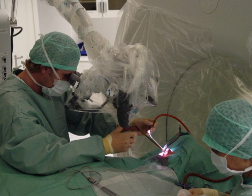
Background: During trans-sphenoidal microsurgical resection of pituitary adenomas, the extent of resection may be difficult to assess, especially when extensive suprasellar and parasellar growth has occurred. In this prospective study, we investigated whether intra-operative magnetic resonance imaging (iMRI) can facilitate tumour resection.
Methods: Twenty patients with macroadenomas, (16 non-functioning, three growth-hormone secreting and one pharmaco-resistant prolactinoma) were selected for surgery in the iMRI. The mean tumour diameter was 27 mm (range 11-41). The mean parasellar grade, according to the Knosp classification, was 2.3. Pre-operative coronal and sagittal T1-weighted and T2-weighted images were obtained. The trans-sphenoidal tumour resection was performed at the edge of the tunnel of a Signa SP 0.5-Tesla MRI. The surgeon aimed at a radical tumour resection that was followed by a peri-operative MRI scan. When a residual tumour was visualised and deemed resectable, an extended resection was performed, followed by another MRI scan. This procedure was repeated until the imaging results were satisfactory. In all patients, we were able to obtain images to assess the extent of resection and to classify the resection as either total or subtotal.
Results: After primary resection, eight out of 20 cases were classified as total resections. A second resection was performed in 11 of 12 cases classified as subtotal resections, and in four of these, total resection was achieved. A third resection was performed in three of the remaining seven cases with subtotal resections, but we did not achieve total resection in any of these cases. Therefore, the use of iMRI increased the number of patients with total resection from 8/20 (40%) to 12/20 (60%). The only observed complication was a transient spinal fluid leakage.
Conclusion: Intra-operative MRI during trans-sphenoidal microsurgery is useful in selected patients for a safe and more complete resection.
Figures

Comment in
-
The value of intra-operative MRI in trans-sphenoidal pituitary surgery.Acta Neurochir (Wien). 2011 Jul;153(7):1375-6. doi: 10.1007/s00701-011-1005-6. Epub 2011 Apr 27. Acta Neurochir (Wien). 2011. PMID: 21523360 No abstract available.
References
-
- Alameda C, Lucas T, Pineda E, Brito M, Uria JG, Magalon R, Estrada J, Barcelo B. Experience in management of 51 non-functioning pituitary adenomas: indications for post-operative radiotherapy. J Endocrinol Invest. 2005;28(1):18–22. - PubMed
-
- Bohinski RJ, Warnick RE, Gaskill-Shiplev MF, Zuccarello M, van Loveren HR, Kormos DW, Tew JM., Jr Intraoperative magnetic resonance imaging to determine the extent of resection of pituitary macroadenomas during transsphenoidal microsurgery. Neurosurgery. 2001;49(5):1133–1143. - PubMed
Publication types
MeSH terms
LinkOut - more resources
Full Text Sources
Other Literature Sources
Medical
