Case Reports
. 2011 Jul 14;365(2):127-38.
doi: 10.1056/NEJMoa1100066. Epub 2011 Apr 27.

IRF8 mutations and human dendritic-cell immunodeficiency

Affiliations
Case Reports

IRF8 mutations and human dendritic-cell immunodeficiency

Sophie Hambleton et al. N Engl J Med. .

Abstract

Background: The genetic analysis of human primary immunodeficiencies has defined the contribution of specific cell populations and molecular pathways in the host defense against infection. Disseminated infection caused by bacille Calmette-Guérin (BCG) vaccines is an early manifestation of primary immunodeficiencies, such as severe combined immunodeficiency. In many affected persons, the cause of disseminated BCG disease is unexplained.

Methods: We evaluated an infant presenting with features of severe immunodeficiency, including early-onset disseminated BCG disease, who required hematopoietic stem-cell transplantation. We also studied two otherwise healthy subjects with a history of disseminated but curable BCG disease in childhood. We characterized the monocyte and dendritic-cell compartments in these three subjects and sequenced candidate genes in which mutations could plausibly confer susceptibility to BCG disease.

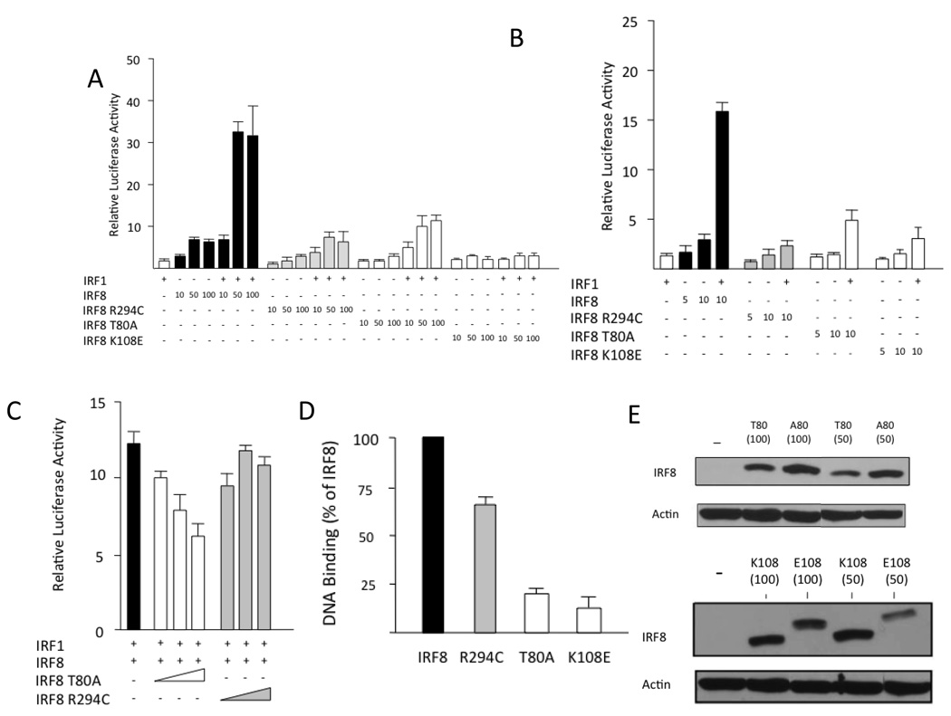
Results: We detected two distinct disease-causing mutations affecting interferon regulatory factor 8 (IRF8). Both K108E and T80A mutations impair IRF8 transcriptional activity by disrupting the interaction between IRF8 and DNA. The K108E variant was associated with an autosomal recessive severe immunodeficiency with a complete lack of circulating monocytes and dendritic cells. The T80A variant was associated with an autosomal dominant, milder immunodeficiency and a selective depletion of CD11c+CD1c+ circulating dendritic cells.

Conclusions: These findings define a class of human primary immunodeficiencies that affect the differentiation of mononuclear phagocytes. They also show that human IRF8 is critical for the development of monocytes and dendritic cells and for antimycobacterial immunity. (Funded by the Medical Research Council and others.).


Figures

Figure 1
Figure 1. Severe depletion of the antigen presenting cell (APC) compartment in autosomal recessive IRF8 deficiency
(A)Flow cytometric evaluation of peripheral blood of affected individual P1 and parents. (B) Flow cytometric evaluation of CD45+HLA-DR+ mononuclear cells from collagenase digested dermis of P1 and control. (C) Quantification of peripheral blood APC subsets as % of mononuclear cells in P1 (red circle), mother (filled circle) and father (open circle) compared with normal adult range (box and whiskers: mean and range; n=28). (D) Quantification of dermal APC subsets as % of live cells, compared with normal adult range (box and whiskers: mean and range; n=22) and Langerhans cells per mm2 compared with normal range (box and whiskers: mean and range; n=12). (E) Affected P1 Langerhans Cells (LC) in an epidermal sheet (×400 magnification; inset × 100), revealed by anti-CD1a immunofluorescence.(F–H) Whole blood cytokine responses measured in vitro. (F) IL-12 production in response to BCG or LPS plus exogenous IFNγ. (G) IFNγ production elicited by indicated stimuli. (H) Production of TNFα, IL-6 and IL-10 in response to indicated combinations of BCG, LPS and exogenous IFNγ. Control, empty bars; affected individual, filled bars; *, undetectable.
Figure 2
Figure 2. Genetic analysis of autosomal recessive (AR) and autosomal dominant (AD) IRF8 deficiency
(A) Schematic representation of the IRF8 gene, including the 9 non-coding (empty) and coding (filled) exons, with the major structural features of the protein shown (DBD, DNA Binding Domain; IAD, IRF Association Domain). The position of the T80A and K108E mutations (bold) in the DBD is shown. (B) Multiple sequence alignment of IRF8 from different species (T80 and K108 are shown in red; stars identify invariant residues). (C) Segregation of K108E in the proband family. (D) T80A is a de novo mutation that has arisen independently in two unrelated MSMD families from Chile and Brazil. (E) Molecular modeling of wild type K108 (panel 1), and T80 (panel 3) and mutant variants E108 (panel 2) and A80 (panel 4) on the three-dimensional structure of DNA-bound IRF8. The hydrogen bond formed between the side chain amino group of K108 and the sugar backbone of DNA is interrupted by E108. The replacement of Thr80 with an Ala residue in the key DNA-binding helix (inserted into the major groove) alters the hydrophobic interface between the protein and the DNA.
Figure 3
Figure 3. Functional characterization of the mutant IRF8 variants
(A, B) RAW macrophages were transiently transfected with various amounts (indicated) of the IRF8 variants, with and without IRF1, and IRF8-dependent transactivation potential was monitored with IL12p40 (A) or iNOS (B) promoter constructs linked to a luciferase reporter gene. (C) The ability of T80A and R294C to suppress transactivation of the iNOS promotor by WT IRF8 was evaluated by co-transfecting RAW macrophages with various amounts of the T80A and R294C variants and fixed amounts of WT IRF8. (D) The relative binding affinity of IRF8 variants for the IL12p40 promoter was estimated by chromatin imunoprecipitation in transfected RAW macrophages. (E) Western blot analysis of cell extracts from RAW macrophages expressing wild-type, or the T80A or the K108E mutant IRF8 variants, with detection based on an HA epitope tag inserted in-frame in all constructs.
Figure 4
Figure 4. Selective depletion of dendritic cells in autosomal dominant IRF8 deficiency
(A) Blood CD1c+ dendritic cells are gated on HLA-DR+ Lin (CD14 CD16 CD19) cells, and expression of CD11c+ and CD1c+ is shown. Percentage of DR+ CD11c+ Lin cells that are positive for CD1c+ is shown for the T80A individuals (P1 ●, P2 ▲), their relatives and travel controls (○,△) (mean—). Numbers on contour plots represent the mean of two independent samples. (B) Blood CD141+ dendritic cells are gated on HLA-DR+ Lin (CD14 CD16 CD19) cells, and expression of CD11c and CD141 is shown. Percentage of DR+ CD11c+ Lin cells that are CD141bright is shown. (C) Plasmacytoid dendritic cells are gated on HLA-DR+ Lin (CD14 CD16) cells, and expression of CD123 and BDCA2 is shown. Percentage of DR+ cells that are positive for CD123 and BDCA2 is shown. (D) Monoyctes are gated on HLA-DR+ Lin (CD2 CD15 CD19 Nkp46) cells, and expression of CD14 and CD16 is shown. Numbers on contour plots represent the percentage of each subset (CD14+, CD14dim and CD14+CD16+) among total monocytes. Percentage of peripheral blood mononuclear cells (PBMC) that are positive for CD14 or CD16 is shown for P1, his relatives and travel control. (E) Production of IL-12p70 (mean ± SD) in response to R848 by FACS-sorted blood CD1c+ (sorted CD1c+ MDC, n=4), PBMCs depleted of CD1c+ DCs (PBMC CD1c+depleted, n=3)) or mock-depleted (PBMC, n=3) from healthy controls, and PBMCs from P1, his relatives and a control collected at the same time (grey bars).

References

    1. Fischer A. Human primary immunodeficiency diseases. Immunity. 2007;27:835–845. - PubMed
    1. Casanova JL, Abel L. Primary immunodeficiencies: a field in its infancy. Science. 2007;317:617–619. - PubMed
    1. Mellman I, Steinman RM. Dendritic cells: specialized and regulated antigen processing machines. Cell. 2001;106:255–258. - PubMed
    1. Geissmann F, Manz MG, Jung S, et al. Development of monocytes, macrophages, and dendritic cells. Science. 2010;327:656–661. - PMC - PubMed
    1. Flanagan RS, Cosio G, Grinstein S. Antimicrobial mechanisms of phagocytes and bacterial evasion strategies. Nat Rev Microbiol. 2009;7:355–366. - PubMed

Publication types

MeSH terms