. 2011 Apr 27;6(1):43.
doi: 10.1186/1748-717X-6-43.

Value of diffusion weighted MR imaging as an early surrogate parameter for evaluation of tumor response to high-dose-rate brachytherapy of colorectal liver metastases

Affiliations

Value of diffusion weighted MR imaging as an early surrogate parameter for evaluation of tumor response to high-dose-rate brachytherapy of colorectal liver metastases

Christian Wybranski et al. Radiat Oncol. .

Abstract

Background: To assess the value of diffusion weighted imaging (DWI) as an early surrogate parameter for treatment response of colorectal liver metastases to image-guided single-fraction ¹⁹²Ir-high-dose-rate brachytherapy (HDR-BT).

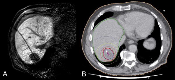
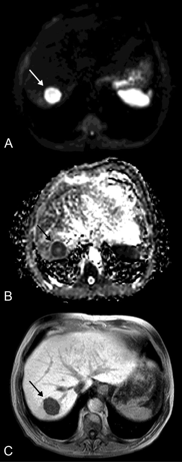
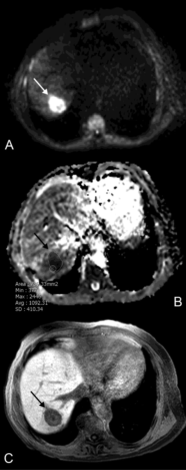
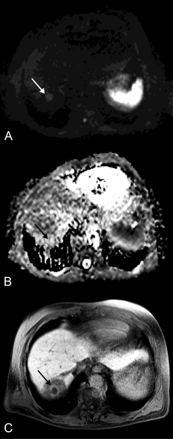
Methods: Thirty patients with a total of 43 metastases underwent CT- or MRI-guided HDR-BT. In 13 of these patients a total of 15 additional lesions were identified, which were not treated at the initial session and served for comparison. Magnetic resonance imaging (MRI) including breathhold echoplanar DWI sequences was performed prior to therapy (baseline MRI), 2 days after HDR-BT (early MRI) as well as after 3 months (follow-up MRI). Tumor volume (TV) and intratumoral apparent diffusion coefficient (ADC) were measured independently by two radiologists. Statistical analysis was performed using univariate comparison, ANOVA and paired t test as well as Pearson's correlation.

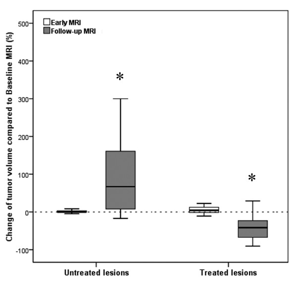
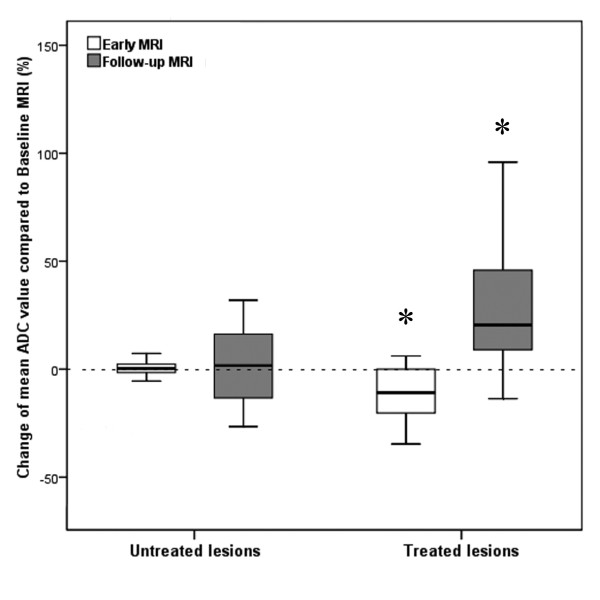
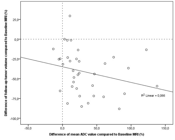
Results: At early MRI no changes of TV and ADC were found for non-treated colorectal liver metastases. In contrast, mean TV of liver lesions treated with HDR-BT increased by 8.8% (p = 0.054) while mean tumor ADC decreased significantly by 11.4% (p < 0.001). At follow-up MRI mean TV of non-treated metastases increased by 50.8% (p = 0.027) without significant change of mean ADC values. In contrast, mean TV of treated lesions decreased by 47.0% (p = 0.026) while the mean ADC increased inversely by 28.6% compared to baseline values (p < 0.001; Pearson's correlation coefficient of r = -0.257; p < 0.001).

Conclusions: DWI is a promising imaging biomarker for early prediction of tumor response in patients with colorectal liver metastases treated with HDR-BT, yet the optimal interval between therapy and early follow-up needs to be elucidated.


Figures

Figure 1
Figure 1
Illustration of MR-guided HDR-BT and 3D dosimetry. 77-year-old man with colorectal liver metastasis in segment VII scheduled for high-dose-rate brachytherapy (HDR-BT). The implantation of one brachytherapy catheter was performed under MRI guidance (A). The tumor enclosing dose (D100) was 21.8 Gy (B).
Figure 2
Figure 2
Baseline MRI preceding HDR-BT. Pre-treatment diffusion-weighted image (DWI) with b = 500 s/mm2 (A), corresponding apparent diffusion coefficient (ADC) map (B) and T1w Gd-EOB-DTPA enhanced MR image in hepatocyte-selective (hepatobiliary) phase (C) of the same patient as in Figure 1 depict the colorectal metastasis in liver segment VII with a mean ADC of 1.29 × 10⁻³ mm2s-1 and a mean volume of 23.3 cm3 (arrow).
Figure 3
Figure 3
Early MRI 3 days after HDR-BT. Early DWI (A) and corresponding ADC map (B) performed 3 days after HDR-BT (same patient as in Figure 1) reveal a decrease in mean ADC by 27.1% to 0.94 × 10⁻³ mm2s-1. The ROI within the lesion indicates an ADC value of 1.09 × 10⁻³ mm2s-1 in this slice of the ADC map (arrow). T1w Gd-EOB-DTPA enhanced MR image in hepatobiliary phase (C) indicates no relevant change in size of the treated lesion (24.1 cm3).
Figure 4
Figure 4
Follow-up MRI. DWI (A) and ADC map (B) performed 105 days post intervention (same patient as in Figure 1) show a rise of mean tumor ADC of 75.2% to 2.26 × 10⁻³ mm2s-1 (arrow). This finding correlates with a decrease in tumor volume by 90.6% (2.2 cm3), depicted in T1w Gd-EOB-DTPA enhanced MR image in hepatobiliary phase (C). The circular hypointense region around the treated lesion in (C) indicates the area of irradiation induced reversible hepatocyte dysfunction.
Figure 5
Figure 5
Boxplot depicting changes of mean volume of non-treated and treated tumors at early and follow-up MRI compared to baseline MRI. Boxplot shows changes of mean tumor volume (TV) of non-treated (*: p = 0.027) and treated colorectal liver metastases (*: p = 0.026) 2 days (early MRI) as well as 3 months (follow-up MRI) after HDR-BT as compared to baseline MRI.
Figure 6
Figure 6
Boxplot depicting changes of mean ADC of non-treated and treated tumors at early and follow-up MRI compared to baseline MRI. Boxplot illustrates changes of mean ADC of non-treated and treated colorectal liver metastases 2 days (early MRI) as well as 3 months (follow-up MRI) following HDR-BT as compared to baseline MRI (*: p < 0.001).
Figure 7
Figure 7
Scatter plot depicting the relationship between changes of mean tumor volume and mean tumor ADC at follow-up MRI compared to baseline MRI. Scatter plot depicts the relationship between changes of mean tumor volumes and mean ADC values of colorectal liver metastases 3 months after treatment with HDR-BT as compared to baseline MRI. A decrease in tumor size is inversely associated with an increase in ADC. Pearson's correlation indicated a weak but statistically significant linear relationship of r = -0.257 (p < 0.001).

References

    1. Rappaport AM. Hepatic blood flow: morphologic aspects and physiologic regulation. Int Rev Physiol. 1980;21:1–63. - PubMed
    1. Kasper HU, Drebber U, Dries V, Dienes HP. [Liver metastases: incidence and histogenesis] Z Gastroenterol. 2005;43:1149–1157. - PubMed
    1. Khatri VP, Petrelli NJ, Belghiti J. Extending the frontiers of surgical therapy for hepatic colorectal metastases: is there a limit? J Clin Oncol. 2005;23:8490–8499. doi: 10.1200/JCO.2004.00.6155. - DOI - PubMed
    1. Guenette JP, Dupuy DE. Radiofrequency ablation of colorectal hepatic metastases. J Surg Oncol. 2010;102:978–987. doi: 10.1002/jso.21658. - DOI - PubMed
    1. Welsh JS, Kennedy AS, Thomadsen B. Selective Internal Radiation Therapy (SIRT) for liver metastases secondary to colorectal adenocarcinoma. Int J Radiat Oncol Biol Phys. 2006;66:S62–S73. - PubMed

Publication types

MeSH terms