. 2011 Jun 17;286(24):21844-52.
doi: 10.1074/jbc.M111.238519. Epub 2011 Apr 27.

Induction of inflammasome-dependent pyroptosis by carbon black nanoparticles

Affiliations

Induction of inflammasome-dependent pyroptosis by carbon black nanoparticles

Anna C Reisetter et al. J Biol Chem. .

Abstract

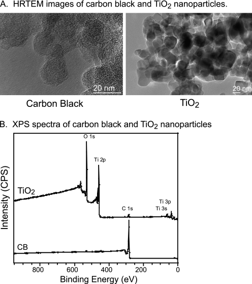
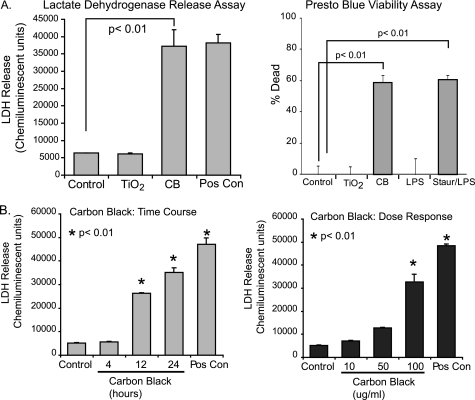
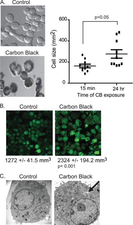
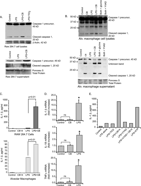
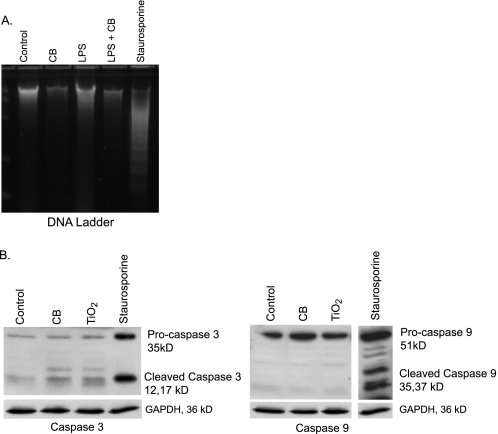
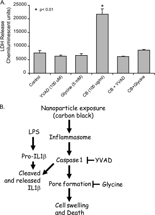
Inhalation of nanoparticles has been implicated in respiratory morbidity and mortality. In particular, carbon black nanoparticles are found in many different environmental exposures. Macrophages take up inhaled nanoparticles and respond via release of inflammatory mediators and in some cases cell death. Based on new data, we propose that exposure of macrophages (both a macrophage cell line and primary human alveolar macrophages) to carbon black nanoparticles induces pyroptosis, an inflammasome-dependent form of cell death. Exposure of macrophages to carbon black nanoparticles resulted in inflammasome activation as defined by cleavage of caspase 1 to its active form and downstream IL-1β release. The cell death that occurred with carbon black nanoparticle exposure was identified as pyroptosis by the protective effect of a caspase 1 inhibitor and a pyroptosis inhibitor. These data demonstrate that carbon black nanoparticle exposure activates caspase 1, increases IL-1β release after LPS priming, and induces the proinflammatory cell death, pyroptosis. The identification of pyroptosis as a cellular response to carbon nanoparticle exposure is novel and relates to environmental and health impacts of carbon-based particulates.


Figures

FIGURE 1.
FIGURE 1.
Nanoparticle characterization. A, shown is high resolution TEM images of CB and TiO2 nanoparticles. The scale bar in both images is 20 nm. Both nanoparticles form aggregates when in solution. Particles were sonicated for 1 h and vortexed vigorously before cell exposure. Primary particle diameters were obtained from the images. B, surface composition of TiO2 and CB nanoparticles was measured using x-ray photoelectron spectroscopy.
FIGURE 2.
FIGURE 2.
CB nanoparticles induce macrophage cell death. A, RAW264.7 cells were exposed to CB (30 μg/cm2) or TiO2 (30 μg/cm2) nanoparticles for 24 h. One group was treated with ATP as a positive control for LDH release. At the end of the culture period, LDH analysis was performed to determine cell viability. The experiment was repeated using identical conditions. A PrestoBlue cell viability assay was performed as indicated by the manufacturer. Significance was determined using nonpaired Student's t test. Staur, staurosporine. B, RAW264.7 cells were exposed to CB nanoparticles (30 μg/cm2) for 4, 12, and 24 h. ATP was used as a positive control for LDH release. At the end of the culture period, an LDH assay was performed to determine cell viability. Additionally, RAW264.7 cells were exposed to CB nanoparticles at 3, 15, or 30 μg/cm2 for 24 h. An LDH assay was performed. Significance was determined using nonpaired Student's t test.
FIGURE 3.
FIGURE 3.
CB nanoparticles cause an increase in macrophage cell size. A, bright field images of CB nanoparticle-exposed cells were obtained 15 min after exposure and again after 24 h. Circles were drawn around representative cells from 10 fields and then used to calculate cellular area. B, fluorescent images of cells were obtained before CB nanoparticle exposure and again 24 h after exposure. Cells were loaded with cell tracker green 5-chloromethylfluorescein diacetate. Average cellular volume was calculated from the level of fluorescence per cell. C, transmission electron microscopy images of control and CB nanoparticle-exposed cells.
FIGURE 4.
FIGURE 4.
CB nanoparticles activate the inflammasome. A, activation of caspase 1 in RAW264.7 cells is shown. 3 × 10⁶ RAW264.7 cells per well were seeded into culture plates. Cells were primed for 3 h with LPS (10 ng/ml). After 3 h medium was aspirated off cells and replaced with fresh medium. LPS was re-added, and CB (30 μg/cm2) or TiO2 (30 μg/cm2) nanoparticles were added for an additional 6 h. After the culture period, whole cell lysates were harvested in Western lysis buffer. Supernatants were concentrated, and proteins were analyzed. Western analysis for caspase 1 was performed on cellular lysates and concentrated supernatants. B, 3 × 10⁶ human alveolar (Alv.) macrophages per well were seeded into culture plates. Cells were primed and cultured as detailed above. Western analysis for caspase 1 was performed on cellular lysates and concentrated supernatants. β-Actin and Ponceau S stain demonstrate equal loading. C, 1 × 10⁶ RAW264.7 cells per well were seeded into culture plates. Cells were cultured as above, except for an extended second incubation time (16–24 h). Supernatants were harvested, and IL-1β levels were measured by ELISA. Significance was determined using nonpaired Student's t test. D, expression of IL-1β, IL-18, and TNFα mRNA in human alveolar macrophages exposed to LPS or CB nanoparticles is shown. 3 × 10⁶ human alveolar macrophages per well were seeded into culture plates. Cells were incubated with CB nanoparticles (30 μg/cm2) or LPS (10 ng/ml) for 4 h. RNA was isolated, and quantitative real-time PCR was performed. *, p < 0.01 by Student's t test (compared with control). ns, not significant. E, 1 × 10⁶ RAW264.7 cells per well were seeded into culture plates. Cells were primed for 3 h with LPS (10 ng/ml). For the second incubation period, LPS was re-added, and CB nanoparticles (30 μg/cm2), TiO2 nanoparticles (30 μg/cm2), YVAD (100 μm), and ATP were added for an additional 24 h. Supernatants were harvested, and IL-1β levels were measured by ELISA.
FIGURE 5.
FIGURE 5.
Nanoparticle-induced cell death is not apoptosis. A, 5 × 10⁶ RAW264.7 cells were seeded into 100-mm culture dishes and incubated with LPS (10 ng/ml), CB nanoparticles (30 μg/cm2), or both for 24 h. One group was exposed to staurosporine (1 μm) as a positive control for apoptosis. After the culture period, DNA was isolated. DNA concentration was measured, and equal concentrations were loaded and run on a SYBR Green gel. The gel was visualized with ultraviolet light, and samples were examined for laddering. B, 3 × 10⁶ RAW264.7 cells/well were seeded into culture plates. Cells were exposed to CB nanoparticles (30 μg/cm2), TiO2 nanoparticles (30 μg/cm2), or staurosporine (1 μm) for 24 h. Proteins were isolated, and Western analysis for the apoptosis-associated caspases 3 and 9 was performed.
FIGURE 6.
FIGURE 6.
Blocking caspase 1 activation protects macrophages from CB nanoparticles toxicity. A, RAW264.7 cells were exposed to CB nanoparticles (30 μg/cm2), CB nanoparticles in combination with the pyroptosis inhibitor glycine (5 mm), CB nanoparticles in combination with the caspase 1 inhibitor YVAD (100 μm), or TiO2 nanoparticles (30 μg/cm2) for 24 h. At the end of the culture period, LDH analysis was performed to determine cell viability. B, the diagram summarizes the CB nanoparticle-induced pathway to inflammasome activation and pyroptosis, as supported by this study.

Comment in

References

    1. Opitz B., van Laak V., Eitel J., Suttorp N. (2010) Am. J. Respir. Crit. Care Med. 181, 1294–1309 - PubMed
    1. Harada R. N., Repine J. E. (1985) Chest 87, 247–252 - PubMed
    1. Twigg H. L., 3rd (2004) Semin. Respir. Crit. Care Med. 25, 21–31 - PubMed
    1. Sibille Y., Reynolds H. Y. (1990) Am. Rev. Respir. Dis. 141, 471–501 - PubMed
    1. Loeffler J., Hedderich R., Fiedeler U., Malsch I., Túquerres G., Koskinen J., Linden M., Lojkowski W., Moritz T., Zins M., Bernabeu E., Larena A. (2010) in Nanoroad SME European Project, European Union, Brussels, Belgium

Publication types

LinkOut - more resources